In a retrospective study, 52 out of 112 leopard geckos had an eye infection such as conjunctivitis. [1] Here’s what you need to know about bacterial conjunctivitis, or pink eye, in leopard geckos.
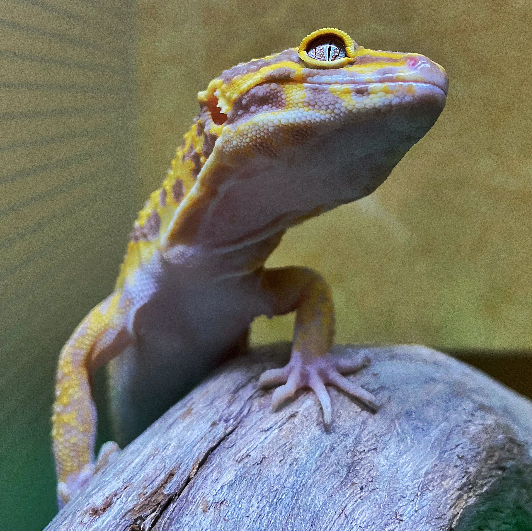
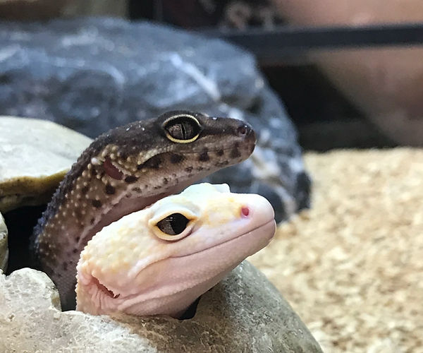
Leopard geckos are popular pocket pets because of their large, colorful eyes. However, captive leopard geckos are extremely likely to develop an eye infection because of their unique eyelid structure. Most geckos have spectacle eyelids, whereas leopard geckos have crinkled eyelids that are fully moveable.
The name “crinkled” comes from the zig-zag pattern of the tissue between the top of the eyelid and the eye’s surface. This eyelid shape creates small pockets where bacteria can easily get trapped and proliferate, causing inflammation to the tissue lining the eyelid, or conjunctiva.
What we’ll cover:
- Symptoms of Conjunctivitis in Leopard Geckos
- Causes of a Leopard Gecko Eye Infection
- Treating Bacterial Conjunctivitis in Leopard Geckos
- [Breaking Case] Fungal Keratoconjunctivitis
What are the symptoms of conjunctivitis in leopard geckos?
Bacterial conjunctivitis is caused by an opportunistic bacterium that has taken advantage of its environment. These bacteria are often commensal species that do not cause harm in small numbers. If there is a vacancy to fill, these bacteria can become problematic as they take up more space.
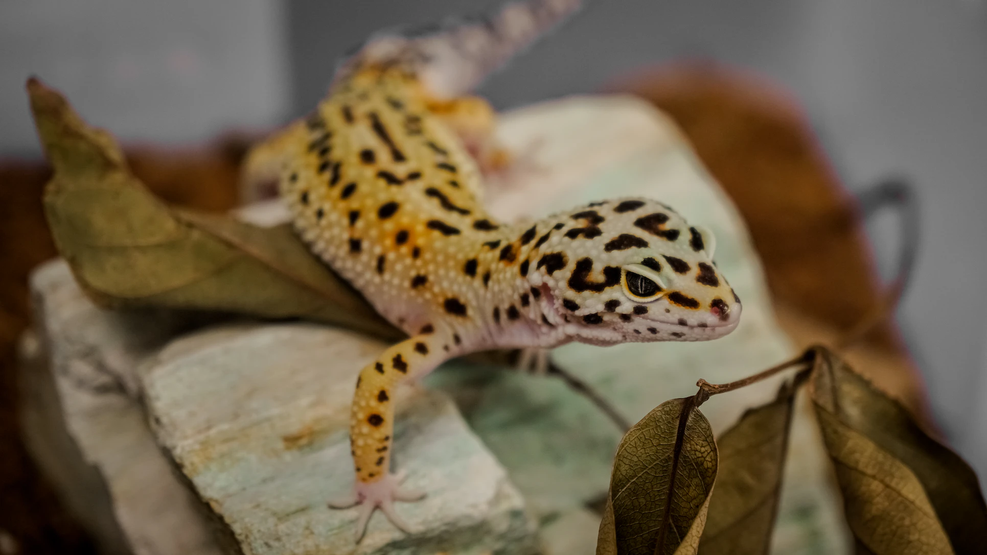
Common signs of conjunctivitis include:
- Not opening eye(s)
- Rubbing eyes
- Eye twitching
- Eye or eyelid swelling
- Pus, discharge, or crust in or around the eye
- Partial or full blindness (visible with corneal haziness)
Left untreated, conjunctivitis can lead to vision loss and septicemia.
What causes a leopard gecko eye infection to develop?
The eyes are an exposed, sensitive organ affected by diet, environment, and eye structure. The crinkled eyelid of leopard geckos leaves them more susceptible to infection; however, the lifestyle of the animal can dramatically impact eye health. Such factors include:
Temperature and humidity
Inappropriate environmental conditions can cause shedding issues, especially around the eyelids.
Improper hygiene
Unclean water sources are breeding grounds for bacteria that can easily come into contact with a gecko’s eye while drinking.
Vitamin A deficiency
Limited evidence suggests that vitamin A deficiency in insectivorous reptiles is common and is linked to epithelial tissue diseases, especially around the eye. [2]
Age
One case series suggests a positive correlation between eye infections and age. [1]
Sex
Female leopard geckos are less likely to develop an eye infection. [1]
Treating bacterial conjunctivitis in leopard geckos
Antibiotic eye drops are a common treatment for conjunctivitis; however, they must be chosen wisely. Using a broad-range antibiotic can worsen the condition by removing the healthy bacteria from the eye. Thus, creating a vacancy for another opportunistic pathogen to fill. It is vital to choose an antibiotic specific to the overgrown, opportunistic pathogen and limit the removal of commensal species.
However, conjunctivitis is a non-specific infection. Many bacteria can become an issue when given the space and nutrients to dominate the microbiome.
So, how do you know which antibiotic will be effective? Identifying what bacteria is causing inflammation is the first step in determining which antibiotic to use. However, there are some obstacles to identification techniques:
1. You need to know its growth requirements for culture.
Each microbe is different and requires different nutrients and temperatures to grow. Most microorganisms are unculturable because their preferred environments can’t be mimicked.
2. Cell counts are vital in determining if an organism is dominating the microbiome.
Understanding, if a sample is out of ten cells or ten million cells, gives the necessary context of the microbes present. Quantification is limited in culture testing and not possible with PCR panels.
3. The bacterium could be an overgrown commensal, not just a pathogen.
Casting a wide net to catch all bacterial species will leave no stone unturned. Panels are limited and require guesswork in choosing which species to test.
Luckily, there is a technique not limited by these obstacles that can confidently detect and quantify all bacterial species present. Microbial DNA sequencing with the MiDOG All-in-One Test allows a snapshot of the microbiome at the moment the sample is taken. No guesswork is needed for the antibiotic profiling of the species of interest either. The MiDOG test includes antibiotic resistance profiling on 43 antibiotics, which informs the microbes’ antibiotic susceptibility.
Breaking case of fungal keratoconjunctivitis
Conjunctivitis is not limited to a bacterial infection; fungi can also cause it. Back in 2019, a six-year-old leopard gecko had keratoconjunctivitis caused by Acremonium and Trichosporon fungal species. It was the first recorded case of fungal keratitis in reptiles caused by these fungi. [3]
Fungal cultures can take up to 3-4 weeks because of slow fungal growth rates, and are 49% more likely to report “no growth” compared to DNA sequencing. [4] A leopard gecko eye infection should be diagnosed and treated as soon as possible to reduce the risk of vision loss or septicemia. MiDOG DNA sequencing detects both bacteria and fungi and has a faster turnaround time of 2-4 business days.
If you think your leopard gecko patient may be suffering from conjunctivitis, consider using the MiDOG test to determine confidently and quickly.
Find out if your vet uses MiDOG before you book your next appointment!
References
[1] Wiggans, K. et.al. (2018). Diagnosis, treatment, and outcome of and risk factors for ophthalmic disease in leopard geckos (Eublepharis macularius) at a veterinary teaching hospital: 52 cases (1985– 2013). JAVMA, 252(3). https://doi.org/10.2460/javma.252.3.316.
[2] Boyer, T. (2018). Vitamin A deficiency in insectivorous lizards. Clinician’s Brief. https://www.cliniciansbrief.com/article/vitamin-deficiency-insectivorous-lizards.
[3] Munevar, C. et.al. (2019). Acremonium and trichosporon fungal keratoconjunctivitis in a Leopard Gecko (Eublepharis macularius). Veterinary Ophthalmology, 22(6), 928. https://doi.org/10.1111/vop.12700.
[4] Damerum, A. et.al. (2023). Next-generation DNA sequencing offers diagnostic advantages over traditional culture testing. AJVR, 84(8). https://doi.org/10.2460/ajvr.23.03.0054.