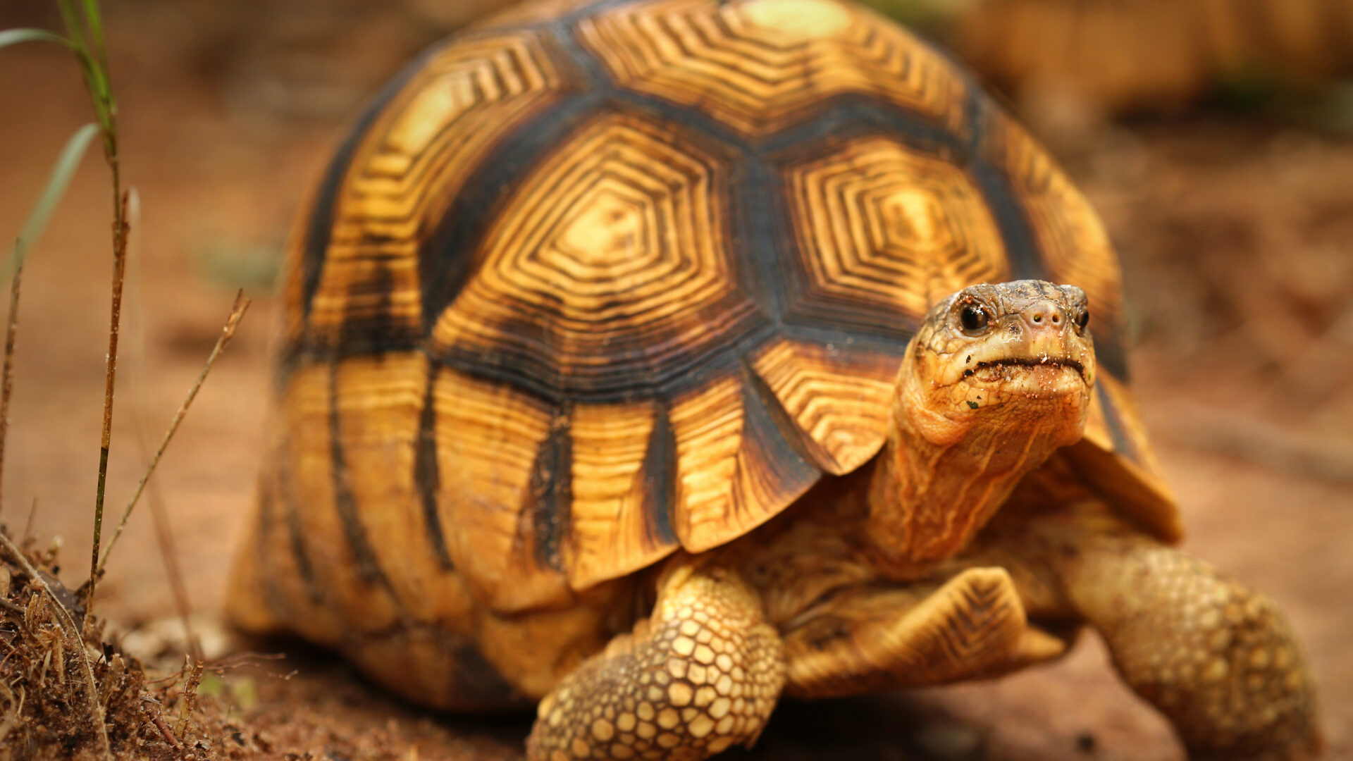
While you might not expect a tortoise to be running anywhere, that doesn’t mean they don’t get runny noses! Tortoise mycoplasmosis is one of the most common diseases in tortoises and is the main cause of upper respiratory tract disease (URTD) in these slow-placed reptiles [1]. Therefore, a combination of understanding the conditions that cause the infection and the infective pathogens themselves is critical in delivering quality healthcare to your reptile.
URTDs are uncomfortable at best for your scaly friend, and if left untreated may result in serious health complications, and in severe cases even death. Tortoises are often symptomatic only when the disease is advanced, and even when diagnosed, treatment can be difficult if the strains of bacteria present are antibiotic-resistant [2]. If you suspect your tortoise has an upper respiratory infection, it is recommended that you make an appointment with your veterinarian to diagnose and provide a treatment plan.
What is Tortoise Mycoplasmosis?
Mycoplasmosis is common in tortoises and is a respiratory disease that can go on to cause secondary URTDs by creating an optimal environment for opportunistic bacteria to proliferate [3]. Mycoplasmosis and subsequent URTDs are pathogen load-dependent and associated with increased levels of M. agassizii and/or M. testudineum [2]. Typically the nasal lesions caused by M. testudineum are less severe than that of M. agassizii, and current studies are assessing other possible Mycoplasma species that may be causing URTDs in tortoises, although research is limited [2]. A study characterizing the bacteria associated with URTDs found that tortoises inflicted with the infectious disease were more likely to have Gram-negative bacteria in their nasal cavity than healthy tortoises, suggesting that mycoplasmosis makes tortoises more susceptible to secondary URTDs [4].
Considering that mycoplasmosis typically presents as a URTD, the disease normally impacts the nasal cavity, although pneumonia does occur. Lesions occur in the nasal cavity and can present in distinct locations or diffusely, with ranging severity [2]. Increased immune responses with basal cell hyperplasia and lymphoid hyperplasia may occur as well [2]. Clinical classification of mycoplasmosis lesions is broken down into three categories: 1) mild inflammation, 2) moderate inflammation, and 3) severe inflammation [2].
For any tortoise, identifying symptoms of mycoplasmosis is critical. Symptoms include:
- Increased mucosal drainage from mouth and nose
- Ocular discharge
- Noninflammatory eyelid edema
- Difficulty breathing
- Decreased appetite
- Excessive sneezing, coughing, or gasping
This disease may progress slowly and can take months or years for a tortoise’s immune system to mount a proper adaptive immune defense against Mycoplasma spp. [3]. Consequently, inflammatory responses to mycoplasmosis occur faster than antibody production and so it is not uncommon for the clinical presentation of the disease to occur without pathogens being detected. Subclinical infections of Mycoplasma spp. can occur, and so understanding the severity of the infection and the possibility of recurrence is key in preventing the disease from advancing to clinical stages [4]. Comorbid tortoises with mycoplasmosis may suffer severe physical wasting and multisystemic organ failure [4].
Prevention and Treatment of Tortoise Mycoplasmosis
As the Manual of Exotic Pet Practice puts it, “the majority of diseases observed in captive reptiles are directly associated with improper husbandry” [5]. This holds true for tortoise mycoplasmosis manifestation, as Mycoplasma spp. proliferation most often occurs following environmental stresses that either directly impact the tortoise’s immune system or cause a shift in the diversity of microbiota in the nasal cavity [4].
Considering the serious nature of mycoplasmosis, it is important to consult with a veterinarian as soon as possible. Delay in care is painful and may result in further medical complications and even death. While treatment will likely entail injectable antibiotics, your veterinarian may also recommend administering nasal drops. Special attention should be given to wiping away nasal discharge to prevent nasal blockages. Treatment should result in improvement within a few days, but there is no guarantee that antibiotics will eliminate the opportunistic Mycoplasma spp., as recurrence is likely [4].
Your veterinarian will also help you identify possible lifestyle changes you and your reptile can make to improve their quality of life and lessen the risk of recurrent infections. This entails understanding the exact pathogen that is impacting your reptile, with modern technological advances allowing for more targeted clinical diagnostic interventions.
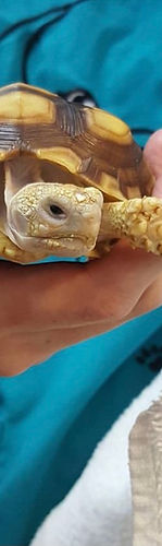
Sultana tortoise making a visit to an exotic pet veterinarian.
Diagnosing Tortoise Mycoplasmosis
With the rise of antibiotic-resistant strains of pathogens, advances in recent clinical diagnostics have allowed for more targeted and efficient interventions. Historically, culture-based methods have been used to assess the nasal microbiome of reptiles, but there are notable diagnostic shortcomings. Specifically, M. agassizii is a small, aerobic, and slow-growing species that are difficult to culture and produces numerous false negatives [6]. Recent studies have begun to shift away from problematic liquid cultures (that require multiple weeks of isolation) in favor of metagenomic sequencing [6,7,8].
Next-Gen Sequencing (NGS) has increasingly helped researchers and veterinarians characterize tortoise mycoplasmosis. A recent study assessing infectious diseases in tortoises has also affirmed the applicability of NGS technology in characterizing Mycoplasma spp. and URTD-associated pathogens [8]. This indicates the clinical applicability of using genomic sequencing to identify, analyze, and eventually treat reptiles more effectively.
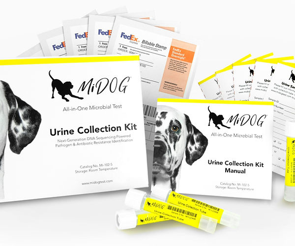
Despite its name, the MiDOG All-in-One Microbial Test offers comprehensive diagnostic information for tortoises suffering from mycoplasmosis. Utilizing NGS technology to detect and quantify all microbial DNA through untargeted and comprehensive sequencing and quantitative comparisons to reference databases, the MiDOG NGS technology provides a useful opportunity to shed light on the microbial makeup of your reptile’s infection for clinical application. The MiDOG microbiome test is a microbial identification test grounded on scientific research that provides veterinarians DNA evidence for the guided treatment of tortoise infections, such as mycoplasmosis and URTDs.
Learn more tips in this extensive guide to tortoise care.
Find out if your vet uses MiDOG before you book your next appointment!
For health-related questions about your tortoise or other exotic pets, reach out to a veterinarian that specializes in exotic pets.
References:
[1] Goessling, J., Guyer, C., Godwin, J., Hermann, S., Sandmeier, F., Smith, L., & Mendonça, M. (2019). Upper respiratory tract disease and associated diagnostic tests of mycoplasmosis in Alabama populations of Gopher tortoises, Gopherus polyphemus. PLOS ONE, 14(4), e0214845. doi: 10.1371/journal.pone.0214845
[2] Jacobson, E., Brown, M., Wendland, L., Brown, D., Klein, P., Christopher, M., & Berry, K. (2014). Mycoplasmosis and upper respiratory tract disease of tortoises: A review and update. The Veterinary Journal, 201(3), 257-264. doi: 10.1016/j.tvjl.2014.05.039
[3] Aiello, C., Esque, T., Nussear, K., Emblidge, P., & Hudson, P. (2018). The slow dynamics of mycoplasma infections in a tortoise host reveal heterogeneity pertinent to pathogen transmission and monitoring. Epidemiology And Infection, 147. doi: 10.1017/s0950268818002613
[4] Brown, M., Brown, D., Klein, P., McLaughlin, G., Schumacher, I., & Jacobson, E. et al. (2001). Mycoplasma agassizii sp. nov., isolated from the upper respiratory tract of the desert tortoise (Gopherus agassizii) and the gopher tortoise (Gopherus polyphemus). International Journal Of Systematic And Evolutionary Microbiology, 51(2), 413-418. doi: 10.1099/00207713-51-2-413
[5] Deczm, M. M. D. M. P. & Tully Jr. DVM MS DABVP (Avian) DECZM (Avian), Thomas N. (2008). Manual of Exotic Pet Practice (1st ed.). Saunders.
[6] Luzuriaga-Neira, A., Sandmeier, F., Weitzman, C., Tracy, C., Bauschlicher, S., Tillett, R., & Alvarez-Ponce, D. (2021). Mycoplasma agassizii, an opportunistic pathogen of tortoises, shows very little genetic variation across the Mojave and Sonoran Deserts. PLOS ONE, 16(2), e0245895. doi: 10.1371/journal.pone.0245895
[7] Weitzman, C., Sandmeier, F., & Tracy, C. (2018). Host species, pathogens and disease associated with divergent nasal microbial communities in tortoises. Royal Society Open Science, 5(10), 181068. doi: 10.1098/rsos.181068
[8] Elbers, J., Brown, M., & Taylor, S. (2018). Identifying genome-wide immune gene variation underlying infectious disease in wildlife populations – a next generation sequencing approach in the gopher tortoise. BMC Genomics, 19(1). doi: 10.1186/s12864-018-4452-0
Categories: Exotic Pets, Next-Gen DNA Sequencing Technology, Reptiles/Amphibians, Respiratory Infection, Turtles