
When Skin Cases Won’t Quit: What Next-Generation Sequencing Revealed in Three Difficult Patients
Every dermatology practice has them: the infection that keeps coming back, the culture that grows nothing useful, the lesion that ignores every reasonable treatment. In a recent MiDOG webinar, board-certified veterinary dermatologist Dr. Jeff Tinsley of the Animal Dermatology Clinic in Louisville, Kentucky, walked through three of his own real-world cases where next-generation sequencing (NGS) helped crack the problem.
MiDOG’s test is billed as an “all-in-one” panel. From a single sample (skin, ear, nasal swab, urine, or feces), it uses DNA sequencing to identify bacteria, fungi, and now parasites, along with virulence genes and antimicrobial resistance markers. Turnaround is about 48 hours once the sample reaches the lab, and samples are stable at room temperature.
Dr. Tinsley uses it most for atypical bacterial infections and rare fungal infections. Here’s what he found.
Watch the Webinar Here
Start with the pattern
Dr. Tinsley sorts tricky cases into two broad camps. If the itch comes first and the skin looks normal until the patient starts licking and scratching, he leans toward an allergy workup.
If the case doesn’t fit allergy, there are usually spontaneous lesions. Those point toward infectious, endocrine, genetic, neoplastic, or autoimmune disease. All three patients below fell into this second camp, and all three had some degree of immune compromise.
Case 1: Duncan, the Boxer with a hidden Nocardia infection
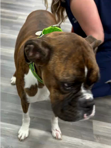
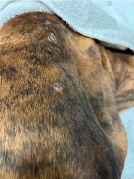
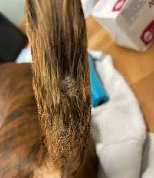
Duncan, a 3-year-old neutered male Boxer, was referred from neurology. He was on cyclosporine for steroid-responsive meningitis-arteritis, had recently come off prednisone, and had a long list of other medications. He arrived with small, gray, crusted plaques scattered across his body.
Cytology showed cocci with nuclear streaming, a sign of active infection, and culture grew a highly resistant MRSP. Chlorhexidine shampoo and mousse, followed by chloramphenicol, cleared those lesions. A first round of biopsies showed only the expected bacterial infection.
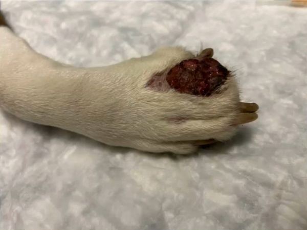
Then, as neurology began tapering his cyclosporine, one of Duncan’s toes started to swell. By his eight-week recheck, it had become a 5 to 6 cm ulcerated, cavitated lesion. Dr. Tinsley’s differentials now included neoplasia, severe demodicosis, and deep fungal or atypical bacterial infection.
What MiDOG found
Dr. Tinsley took new biopsies and, before closing, swabbed deep inside the punch site for MiDOG. The report showed Nocardia making up 98.2% of the bacteria in the sample.
Histopathology described pyogranulomatous dermatitis with filamentous, variably acid-fast bacteria. After Dr. Tinsley shared the MiDOG report with the pathologist, special stains confirmed Nocardia. As he pointed out, stains only work if the biopsy happens to capture the organisms, so knowing what to look for helps.
Treatment and outcome
A surgeon debulked the lesion and sent tissue to Texas A&M for culture, with a heads-up to look for atypical organisms. The lab confirmed Nocardia, found a methicillin-resistant staph, and also grew Scedosporium, an environmental fungus that rarely causes disease.
Duncan was treated with doxycycline, trimethoprim-sulfa, and terbinafine. The sulfa drug was stopped after it triggered hypothyroidism and mild anemia, both of which resolved. He continued doxycycline for about four more months and terbinafine for about three.
At eight months, Duncan had no new lesions and was off all antimicrobials, prednisone, and cyclosporine. About a year and a half after his first visit, he was still doing excellently.
One caution from Dr. Tinsley: MiDOG’s resistance chart is not a culture and sensitivity, and for an uncommon organism like Nocardia, it may have limited antibiotic data. You may still need additional research or testing to choose a drug.
View the Report Here
Case 2: Ariana, the Shih Tzu mix with a rare fungus
Ariana, an 11-year-old spayed female Shih Tzu mix, was being treated by neurology for meningoencephalitis of unknown origin. She had been on prednisone for over a year, plus a long course of cyclosporine, and her bloodwork showed markedly elevated liver enzymes.
She came in with two problems: firm, thickened, hairless lesions spreading up her right elbow, and a large, granular, cellulitis-like mass on a front toe that had been growing for about five months.
Reading the report closely
The elbow biopsy showed calcinosis cutis, likely a consequence of the long-term steroids. The toe biopsy showed deep fungal hyphae.
At first glance, the MiDOG report seemed to say nothing was there: no known bacterial or fungal pathogens were flagged. But Dr. Tinsley looked at the microbial overview. The sample was 100% fungal, with no bacteria at all, and Paraconiothyrium made up 97% of it.
This is a key lesson from the webinar. A “no pathogens detected” summary doesn’t always mean nothing is going on, especially for organisms with little published data. Dr. Tinsley researched the organisms listed, focused on the most abundant one, and shared the report with the dermatopathologist, who agreed the findings fit.
Paraconiothyrium belongs to the hyalohyphomycoses, a group of environmental fungi that can cause opportunistic skin and paw infections in immunosuppressed animals.
Treatment and outcome
Neurology tapered her steroids, and the affected toe was amputated. Dr. Tinsley recommended sending the amputated tissue for pathology too, since large, thickened lesions can hide something walled off in the center. Terbinafine was started but stopped after GI upset and tremors.
Her original skin lesions resolved. Sadly, Ariana was later euthanized, believed to be due to a flare of her neurologic disease. But she had a significantly longer stretch of comfortable skin.
Case 3: Taco, the cat with a surprise finding
Taco, a 5-year-old neutered male domestic shorthair, had a two-month history of one swollen central paw pad. It wasn’t painful or itchy, and it hadn’t responded to prednisolone. He is also FIV-positive.
Dr. Tinsley’s short list included plasma cell pododermatitis, eosinophilic granuloma, a deep or atypical infection, and, less likely, neoplasia. He biopsied the pad and swabbed inside the punch site for MiDOG.
Two answers instead of one
Pathology confirmed plasma cell pododermatitis, the first time Dr. Tinsley had seen it affect a single paw. He fully expected MiDOG to come back clean.
Instead, it found a hemotropic Mycoplasma (likely Candidatus Mycoplasma haemominutum), making up 100% of the organisms detected. These organisms are usually associated with infectious anemia, and their role in skin disease is barely documented. The resistance panel flagged many resistance genes but suggested doxycycline could be effective.
Where things stand
Taco started a slow dexamethasone taper at around 0.3 mg/kg, with some reduction in swelling after two weeks. Doxycycline has just been added for a two- to three-month trial.
Open questions remain. Did his FIV status make him more susceptible to Mycoplasma? Is the Mycoplasma driving the plasma cell pododermatitis, or are they two separate conditions? Dr. Tinsley noted that if Taco fully resolves, the case may be worth reporting in the literature, and he might avoid lifelong immunosuppressive drugs.
Practical tips from Dr. Tinsley
- Sample deep for deep problems. When hunting for rare or atypical organisms, Dr. Tinsley collects sterilely and swabs inside the punch biopsy site before closing, so surface bacteria don’t muddy the result. For extra coverage, he sometimes adds a small piece of sterile tissue, epidermis trimmed off, to the same MiDOG tube.
- Share the report with your pathologist and culture lab. In Duncan’s case, the MiDOG result led to targeted special stains and told the culture lab what to look for.
- Read past the summary. Check the microbial overview and organism breakdown, not just the pathogen flags, as Ariana’s case showed.
- Interpret in context. Weigh every result against the history, physical exam, histopathology, and response to treatment. Some detected organisms are commensals not worth chasing.
- Remember it isn’t a culture. NGS can replace or complement culture and sensitivity in the right cases, but resistance data may be incomplete for uncommon organisms.
- Use it to streamline. One sample can stand in for separate aerobic, anaerobic, fungal, and pythiosis tests, which can save clients money and time, especially for slow-growing organisms like Nocardia.
- Collect now, decide later. Samples in the MiDOG buffer are stable at room temperature for a long time (Dr. Tinsley cited up to a year), so you can collect at biopsy and submit later if cost is a concern.
The bottom line
Across all three cases, NGS didn’t replace clinical reasoning. It sharpened it. In Duncan, it pointed to Nocardia quickly. In Ariana, it identified a fungus few clinicians have heard of. In Taco, it turned up an organism no one was looking for.
As Dr. Tinsley summarized, MiDOG is an excellent choice for suspected deep, rare, and fungal infections, best used with good case selection and alongside, not instead of, the rest of the workup.
These are three individual cases rather than controlled studies, and the webinar was hosted by MiDOG. Dr. Tinsley reported no conflicts of interest. Watch the full webinar.