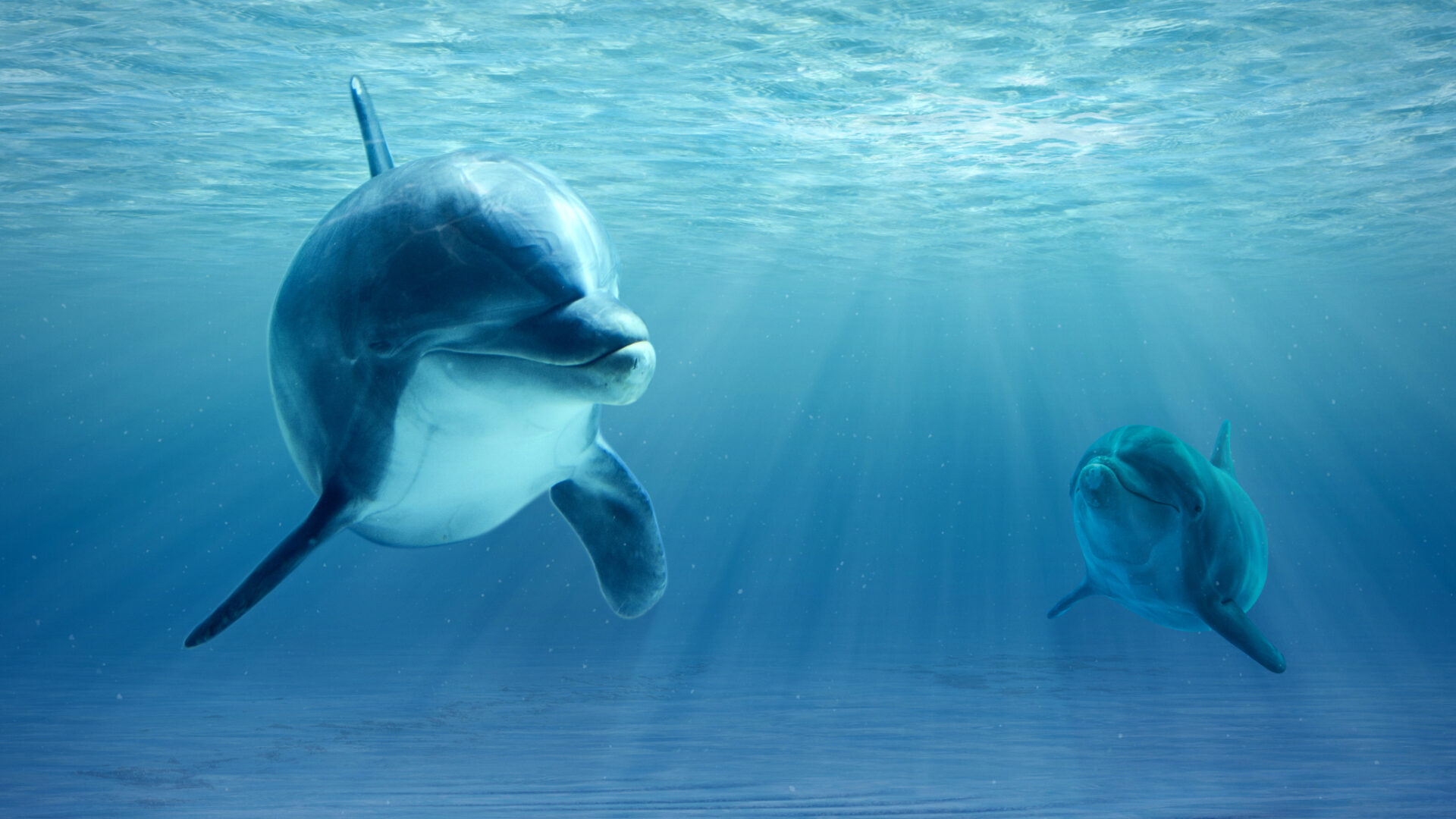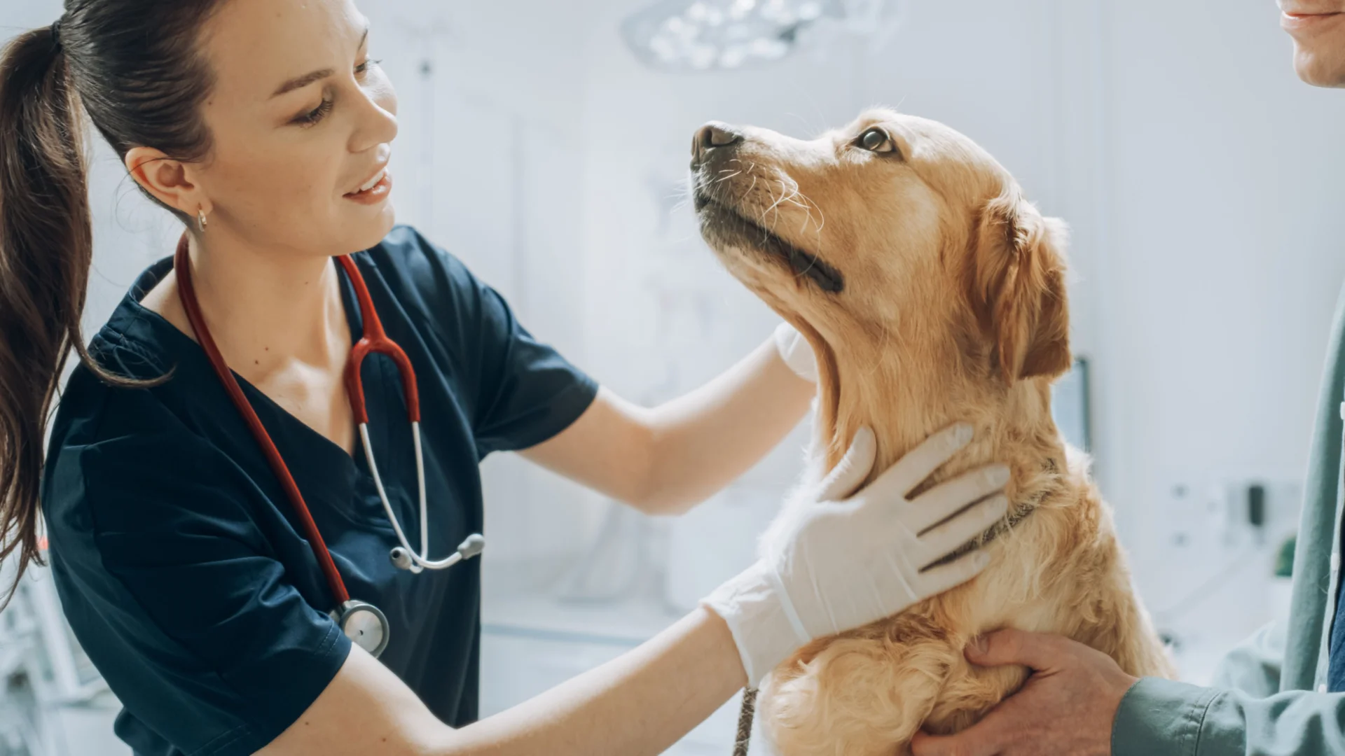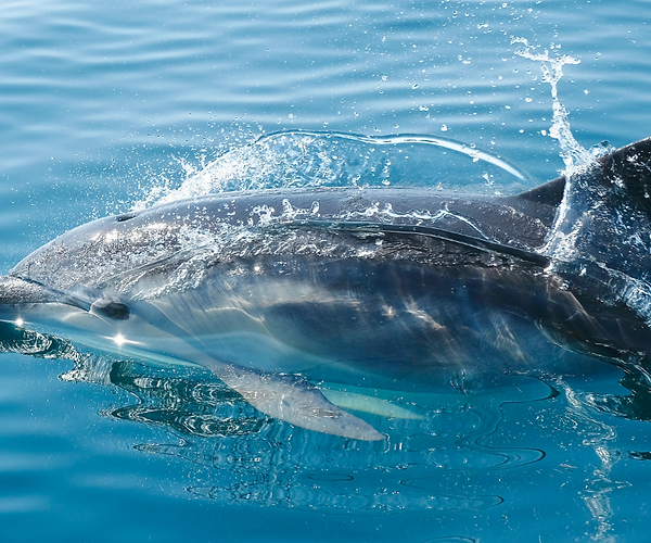
Like humans and other vertebrate species, marine mammals are prone to a variety of ailments, including gastrointestinal (GI) infections, which sometimes occur when environmental stressors compromise the immune system. These charismatic megafauna, especially dolphins, have been well studied because of their presence in managed care facilities and wild stocks worldwide.
Like most wild species, marine mammals are skilled at masking disease signs to prevent predation and help prolong survival. In fact, wild marine mammals will often beach or strand themselves when battling the end stages of disease to avoid predation.
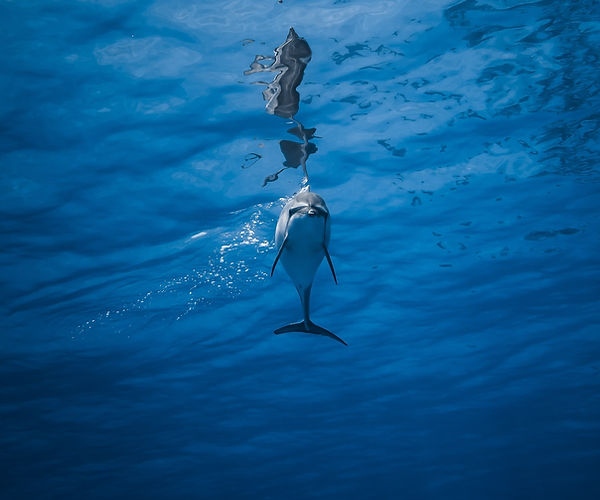
Conversely, when in dolphin captivity these creatures may show subtle illness signs when they have a medical condition, such as a dolphin GI infection. Early detection of GI infections, particularly before clinical signs become obvious, can give cetaceans the
best chance of recovery and survival. A proactive healthcare program utilizing the right healthcare services, including testing for infections and environmental contamination, is essential.
Fortunately, novel molecular-based technologies based on DNA sequencing can help on both fronts.
Common Causes of Gastrointestinal Infections in Dolphins
The dolphin GI physiology is quite unique in that it is similar to the GI system of terrestrial ungulates such as hippos and camels. Their stomachs are separated into three chambers—the forestomach, the fundus, and the pylorus—which allow them to digest their fish-based diet and demineralize bone. Examination of the lower chambers can only be achieved by advanced imaging or post-mortem exam. However, the forestomach may be examined using endoscopy.
Dolphins are prone to a variety of infections, and some may lead to ulceration of the stomach mucosa. One study showed that 17% of wild dolphin carcasses had gastric ulcers, making this a common and concerning problem [1]. Interestingly, it is theorized that stomach ulcers in dolphins may stem from social stressors, similar to ulcers in humans.
In some cases, dolphins may serve as carriers for zoonotic diseases that can also put animal handlers at risk, so it is important to know which pathogens may be present.
Common GI infections that affect dolphins include:
- Bacterial infections — The dolphin GI tract houses a variety of bacteria, including Bacteroides, E. coli, and Enterococcus species [2]. Bacteria reported to cause gastric infections include Clostridium, Pasteurella, Salmonella, Helicobacter, and Vibrio species [3]. However, the presence of these bacterial species does not necessarily mean a marine mammal is suffering from gastritis because some types, including Vibrio, have been cultured from the colon of healthy dolphins [4].
- Fungal infections — Captive marine mammals are especially prone to fungal disease due to concentrated exposure in their enclosures. Because dolphins lack nasal turbinates, which would normally filter out pathogens, fungi can more easily enter their bodies. Candida spp. can be a normal finding in dolphins; however, it has also been associated with GI ulcers [3].
- Viral infections — Although less common than bacterial infections, viral infections can also cause gastrointestinal infections in dolphins and other marine mammals. Norovirus, a common human GI pathogen, has been reported in California sea lions and a harbor porpoise [4]. Additionally, adenovirus has also been reported in captive dolphins [5].
- Parasites — Parasitic nematodes, such as anisakis, are commonly found in beached dolphins and can cause ulcerative gastritis and inflammation.
Gastrointestinal Infection Signs in Dolphins
Dolphins are skilled at masking disease signs, and unfortunately, acute illness and sudden death are often the first signs of a medical problem. This makes timely and accurate diagnosis of potential problems essential.
Identifying dolphin GI problems can be challenging. For example, vomitus can easily be washed away or diluted if not immediately observed. However, subtle signs, such as the presence of fish bones on the bottom of a dolphin captivity pool, could indicate a GI infection, since alterations in the gastric pH can prevent demineralization.
Other subtle signs of dolphin GI infections that handlers should watch for include:
- Decreased or lack of appetite
- Swimming slowly
- Regurgitation or vomiting
- Changes in feces color, consistency, or frequency
- Slight body arching
- Behavioral changes
- Withdrawal from animal care staff
- Weight loss
- Tensed appearance
- Abdominal pain
- Changes in chuff odor
- Beaching, in wild marine mammals
Diagnosing Gastrointestinal Infections in Dolphins
Dolphins with suspected GI infections should receive a nose-to-fluke examination to identify any underlying problems or co-infections. Baseline blood work, including a complete blood count and chemistry, urinalysis, and generalized inflammation tests, like fibrinogen and erythrocyte sedimentation rates (ESR), can help provide an overall health assessment. Other common diagnostics include chuff, gastric, and fecal cytology. Fluid and fecal culture and antibiotic sensitivity tests can also help further pinpoint the source of infection and guide treatment choices.
Although extensive research has been conducted in wild and stranded dolphins, new, emerging gastric infections are continuously reported, making it challenging to determine disease etiologies with conventional testing. Fortunately, advanced diagnostics can give handlers and medical teams unprecedented insights into the health of these stoic creatures. MiDOG Animal Diagnostic’s NGS-based infectious disease diagnostics are microbial DNA-sequencing techniques that identify the presence and composition of microbes in patient samples. MiDOG’s sequencing technology has allowed for the rapid identification of pathogenic microbes in marine mammal fecal samples and the selection of healthy donor material for fecal transplants. This testing can allow for the identification of pathogens in samples from sick and seemingly healthy dolphins, which can help head off problems even before they develop and manifest in clinical signs.
Additionally, pool water and other environmental surfaces, including food fish, fish buckets, and pool surfaces, can also be tested to rule out potential sources of infection. NGS technologies can be used to establish an environmental monitoring program to help predict potential pathogenic infections in marine mammal collections. This is especially important because environmental contamination can affect all of the dolphins in a managed care facility.
Treatment Options for Gastrointestinal Infections in Dolphins
Treatment will depend on the underlying causes of a dolphin’s GI infection. In some cases, gastric infections may be self-limiting. However, early diagnosis and treatment provide the best chance of a rapid recovery and may help prevent more severe ulcerative disease.
Treatment for dolphin GI infections may include:
- Antibiotics
- Gastroprotectant medication, such as sucralfate
- H2-blockers
- Antifungal medications
Advanced treatments, such as fecal material transfer (FMT), have been extensively used in agriculture medicine and have recently been reported in pilot whales. This study was conducted jointly with MiDOG Animal Diagnostics, and FMT may be a therapeutic consideration to help rebalance the gut microbiome in dolphins.
How can MiDOG tests help dolphins?
MiDOG’s All-in-One Microbial Test is an exciting addition to conventional diagnostics that can be used as part of an environmental monitoring program to detect potentially pathogenic bacterial species in a dolphin’s environment. The test can also be used to identify problematic bacteria in GI samples as part of a comprehensive wellness program so that infections can be detected promptly, and treatment can be started before the dolphin becomes clinically ill.
Discover the infectious disease diagnostic advantages of MiDOG!
References
- Abollo, E., Fernando, A.L., Gestal, C., Benavente, P. (1998). Long-term recording of gastric ulcers in cetaceans stranded on the Galacian (NW Spain) coast. Diseases of Aquatic Organisms. 32(1). https://www.researchgate.net/publication/13609316_Long-term_recording_of_gastric_ulcers_in_cetaceans_stranded_on_the_Galician_NW_Spain_coast
- Robles-Malagamba, M. J., Walsh, M. T., Ahasan, M. S., Thompson, P., Wells, R. S., Jobin, C., Fodor, A. A., Winglee, K., Waltzek, T. B. (2020) Characterization of the bacterial microbiome among free-ranging bottlenose dolphins (Tursiops truncatus). Heliyon, 6(6). https://www.ncbi.nlm.nih.gov/pmc/articles/PMC7305398/
- Field, C. L. (2022). Mycotic diseases of marine mammals. Merck Veterinary Manual. https://www.merckvetmanual.com/exotic-and-laboratory-animals/marine-mammals/mycotic-diseases-of-marine-mammals
- Gulland, F. M. D., Dierauf, L. A., Whitman, K. L. (2018). CRC Handbook of Marine Mammal Medicine. CRC Press.
- Rubio-Guerri, C., Garcia-Parraga, D., Nieto-Pelegrin, E., Melero, M., Alvaro, T., Valls, M., Crespo, J. L., Sanchez-Vizcaino, J. M. (2015). Novel adenovirus detected in captive bottlenose dolphins (Tursiops truncatus) suffering from self-limiting gastroenteritis. BMC Veterinary Research. 11(53). https://bmcvetres.biomedcentral.com/articles/10.1186/s12917-015-0367-z
Additional resources:
- https://www.ncbi.nlm.nih.gov/pmc/articles/PMC154630/#:~:text=Gastric%20ulcers%20have%20been%20reported,novel%20 urease%2d Positive%20 Helicobacter%20sp.
- https://journals.asm.org/doi/full/10.1128/aem.66.11.4751-4757.2000
- http://intranet.vef.hr/dolphins/radovi/pdf%202017/2017Hrabar_DAO_gastric%20lesions.pdf
- https://pubmed.ncbi.nlm.nih.gov/24492055/
- https://www.ncbi.nlm.nih.gov/pmc/articles/PMC7305398/
Categories: Exotic Pets, Gastrointestinal Health, Marine Mammal
