Who you calling yellow-bellied? It may not be your reptile’s lack of courage, but rather an infection causing the yellow hue on your scaly friend’s skin. This infection goes by several names, including Chrysosporium anamorph of Nannizziopsis vriesii (CANV), Yellow Fungus Disease (YFD), CANV mycosis, and most recently Onygenalean Dermatomycoses [1]. Debate surrounding the name of the disease stems largely from advancements in technology that have revealed several microorganisms can cause this infection, not just one. Semantics aside, YFD is a very serious condition associated with yellow/brown skin crusts that appear randomly on the skin and is the most common fungal disease in captive reptiles.
This condition is painful for your scaly friend and results in death if left untreated. Early intervention is critical for treatment to be impactful, and so if you suspect your pet has YFD, visit an exotic pet veterinarian immediately so they can diagnose and provide a treatment plan.
What is Yellow Fungus Disease?
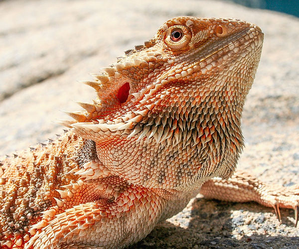
While YFD poses a risk for all reptiles, this infection is the leading cause of mortality in bearded dragons. Additionally, green iguanas, veiled chameleons, Uromastyx, and green water dragons are at a higher risk of contracting YFD as well [2]. This infection is mostly caused by primary, obligate fungi infecting reptiles with weakened immune systems, and is extremely contagious [3].
Specifically, the main pathogens implicated in YFD are fungi from the genera Nannizziopsis, Parannizziopsis, Ophidiomyces, Onygenaceae, Clavicipitaceae, and more [4]. Moreover, Nannizziopsis guarroi are among the fungi implicated as the classical causative agent of YFD in bearded dragons, along with Ophidiomyces ophiodiicola in snakes suffering deep fungal dermatitis [4]. Other obligate pathogenic fungi that may cause YFD belong to the family Clavicipitaceae, which causes granulomatous glossitis, pharyngitis, dermatitis, and visceral mycosis in various reptiles [4]. It is important to note that fungi formerly known as CANV have recently been reassigned to the family Onygenaceae, based on phylogenetic studies using molecular technology [1].
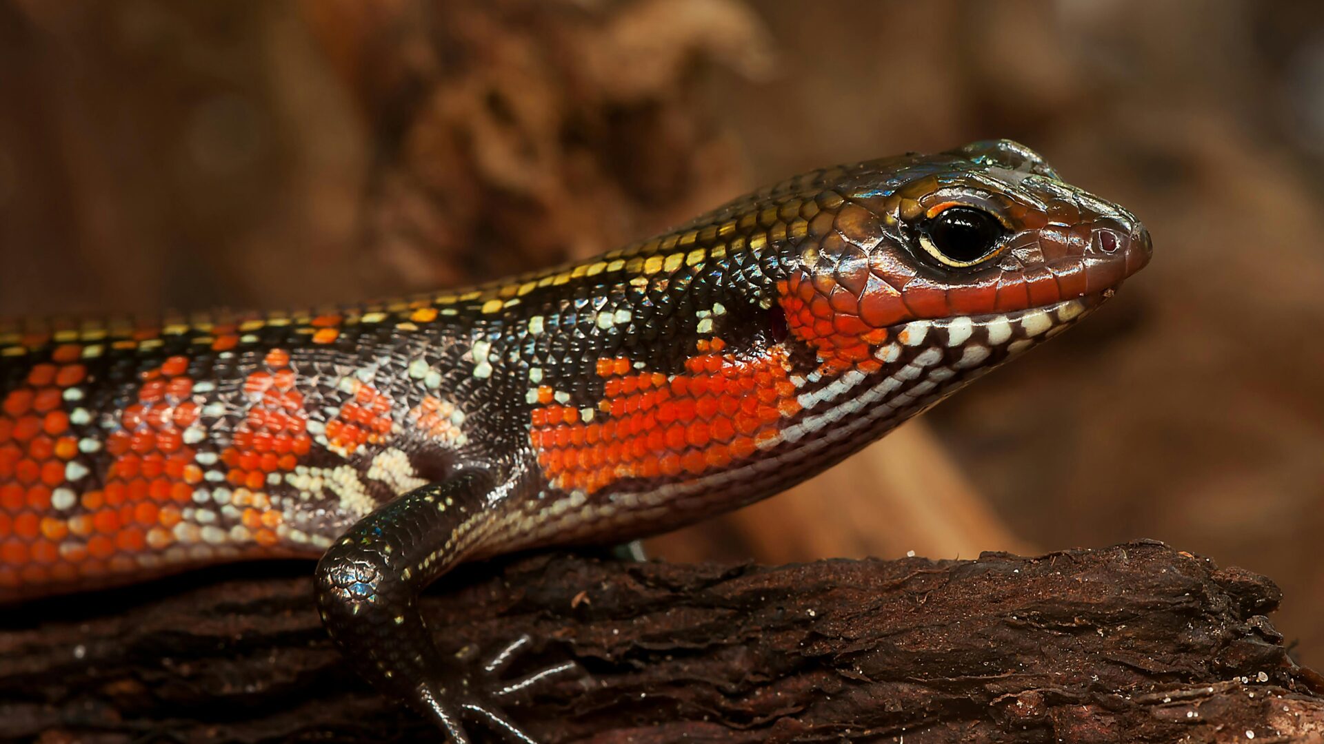
YFD presents with lesions that are similar for all associated fungi, although vary greatly dependent on the host species and environmental factors. These lesions or crusts are initially yellow and then thicken into brown plaques, which may crack and ooze pus or fall off and expose pink dermal inflammation [5]. Lesions may develop over several months from yellow crusts to brown plaques [5]. The location of YFD lesions is most often around the head and mouth, but can occur anywhere on a reptile and can even cover the whole body [6,7]. YFD caused by Nannizziopsis spp. progress slowly, making it difficult for pet owners to identify early warning signs since the disease may not appear debilitating at first [5]. Bearded dragons with YFD are commonly infected with Nannizziopsis spp. [8]. The severity of infection is influenced by the host species, as infection is extremely severe in bearded dragons and often fatal due to the fungal infection spreading to muscle, bone, and internal tissues [8,9].
For any reptile, identifying symptoms of YFD is critical. Symptoms include:
- Yellow/brown lesions or crusts anywhere on the skin that gets larger over time
- Irregular shedding
- Dull or opaque appearance after shedding
- Loss in appetite
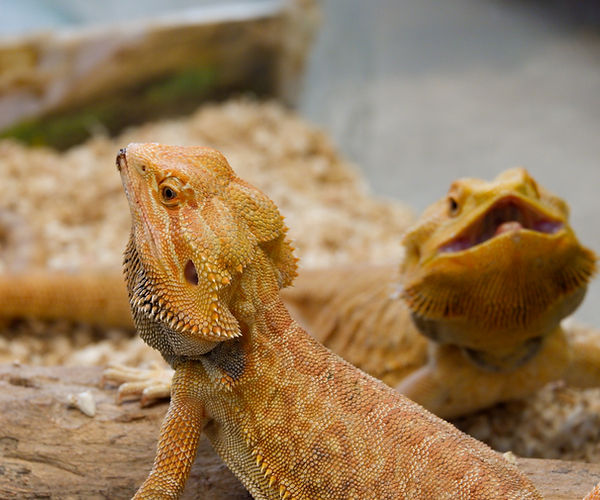
The image above depicts two healthy bearded dragons in a well-maintained terrarium.
Preventing Yellow Fungus Disease in Your Reptile
Pet reptiles are highly at risk for developing fungal infections due to incorrect temperature and humidity levels. As the Manual of Exotic Pet Practice puts it, “the majority of diseases observed in captive reptiles are directly associated with improper husbandry” [10]. Notably, fungal infections in captive reptiles are more common at lower ambient temperatures [5]. In order to prevent Nannizziopsis spp., Paranannizziopsis spp., and O. ophiodiicola infection in reptiles, it is important to reduce the viral load while setting the proper temperature and humidity levels [5]. A proper diet is also very important in maintaining optimal health for your reptile, as vitamin deficiencies can leave your reptile vulnerable to pathogens. And while it may seem obvious that cleaning your pet’s terrarium is important in warding off infections, cleanliness cannot be overstated. Isolating suspected cases of YFD from other reptiles is also necessary.
Treating Yellow Fungus Disease For Reptiles
Considering the serious nature of YFD, it is important to consult with a veterinarian as soon as possible. Delay in care is painful and may result in ineffective treatment. While treatment will likely entail systemic antifungals, topical antifungals, and/or antiseptic solutions, in advanced cases, your reptile may need surgery to remove lesions [9]. Supportive therapy may be necessary for your reptile as well.
Your veterinarian will also help you identify possible lifestyle changes you and your reptile can make to improve their quality of life and lessen the risk of recurrent infections. This entails understanding the exact pathogen that is impacting your reptile, with modern technological advances allowing for more targeted clinical diagnostic interventions.
Diagnosing Yellow Fungus Disease in Reptiles
Historically, culture-based methods have been used to assess the possible fungal infections in reptiles, but there are notable diagnostic shortcomings. In one study comparing the efficacy of culture-based diagnostics with sequencing techniques for reptiles suffering YFD, most isolates except N. vriesii produced “no-growth” cultures or had to be cultured repeatedly to confirm a suspected case [2]. Conversely, using molecular and morphological assessments, the researchers were able to discover six new species in Nannizziopsis and three new species in the new genus Paranannizziopsis [2].
Despite its name, the MiDOG All-in-One Microbial Test may provide the answer to the diagnostic conundrum that YFD poses for your reptile. Utilizing NGS technology to detect and quantify all microbial DNA through untargeted and comprehensive sequencing and quantitative comparisons to reference databases, the MiDOG NGS technology provides a useful opportunity to shed light on the microbial makeup of your reptile’s fungal infection for clinical application. The MiDOG microbial test is grounded on scientific research that provides veterinarians DNA evidence for the guided treatment of reptile infections, such as yellow fungus disease.
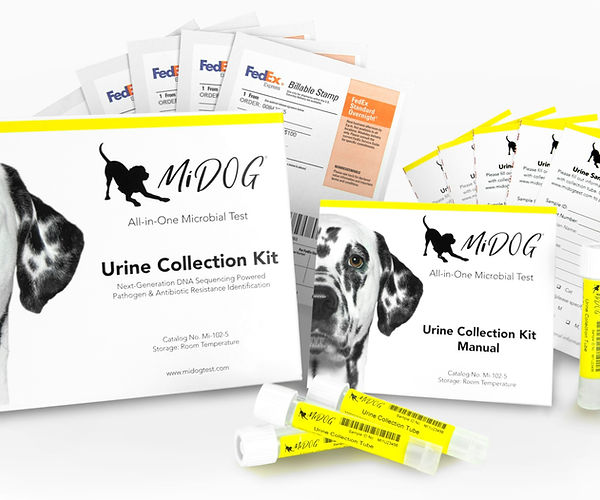
Find out if your vet uses MiDOG before you book your next appointment!
For health-related questions about your reptile or other exotic pet, reach out to a veterinarian that specializes in exotic pets.
References
1. Paré, J., Wellehan, J., Perry, S., Scheelings, T., Keller, K., & Boyer, T. (2021). Onygenalean Dermatomycoses (Formerly Yellow Fungus Disease, Snake Fungal Disease) in Reptiles. Journal Of Herpetological Medicine And Surgery, 30(4). doi: 10.5818/19-12-221.1
2. Sigler, L., Hambleton, S., & Paré, J. A. (2013). Molecular characterization of reptile pathogens currently known as members of the chrysosporium anamorph of Nannizziopsis vriesii complex and relationship with some human-associated isolates. Journal of clinical microbiology, 51(10), 3338–3357. https://doi.org/10.1128/JCM.01465-13
3. Schneider, J., Heydel, T., Klasen, L., Pees, M., Schrödl, W., & Schmidt, V. (2017). Characterization of Nannizziopsis guarroi with genomic and proteomic analysis in three lizard species. Medical Mycology, 56(5), 610-620. doi: 10.1093/mmy/myx083
4. Schmidt, V. (2015). Fungal Infections in Reptiles—An Emerging Problem. Journal Of Exotic Pet Medicine, 24(3), 267-275. doi: 10.1053/j.jepm.2015.06.014
5. Johnson, R., & Sanger, C. (2020). Yellow fungus and related diseases in Australian reptiles. Retrieved 13 July 2021, from https://www.wildlifehealthaustralia.com.au/Portals/0/Documents/FactSheets/Reptiles/Yellow_fungus_disease_in_Australian_reptiles.pdf
6. Paré J, Jacobson E (2007) Mycotic diseases of reptiles. In ‘Infectious diseases and pathology of reptiles.’ (Ed. ER Jacobson.) pp. 527-570. (CRC Press: Boca Raton).
7. Lorch JM, Knowles S, Lankton JS, Michell K, Edwards JL, Kapfer JM, Staffen RA, Wild ER, Schmidt KZ, Ballmann AE (2016) Snake fungal disease: an emerging threat to wild snakes. Philosophical Transactions of the Royal Society B: Biological Sciences 371, 20150457.
8. Bowman MR, Paré JA, Sigler L, Naeser JP, Sladky KK, Hanley CS, Helmer P, Phillips LA, Brower A, Porter R (2007) Deep fungal dermatitis in three inland bearded dragons (Pogona vitticeps) caused by the Chrysosporium anamorph of Nannizziopsis vriesii. Medical Mycology 45, 371-376.
9. Paré J, Sigler L (2016) An overview of reptile fungal pathogens in the genera Nannizziopsis, Paranannizziopsis, and Ophidiomyces. Journal of Herpetological Medicine and Surgery 26, 46-53.
10. Deczm, M. M. D. M. P. & Tully Jr. DVM MS DABVP (Avian) DECZM (Avian), Thomas N. (2008). Manual of Exotic Pet Practice (1st ed.). Saunders.
Categories: Exotic Pets, Fungal Infections, Reptiles/Amphibians, Skin Health, Veterinary Dermatology