MiDOG technology can help diagnose your reptile’s stomatitis infection.
As one of the most common diseases in reptiles, stomatitis affects the oral cavity, tongue, palate, and/or esophagus. More commonly known as mouth rot, stomatitis is associated with inflammation of the mouth consisting of gingivitis, glossitis, palatitis, and cheilitis [1]. While the initial exposure to opportunistic bacteria may be caused by physical trauma within the mouth, environmental factors such as misguided caretaking and nutritional care create the framework for infectious agents to thrive [2]. Therefore, a combination of understanding the conditions that caused the infection and the infective pathogens themselves is critical in delivering quality healthcare to your reptile.
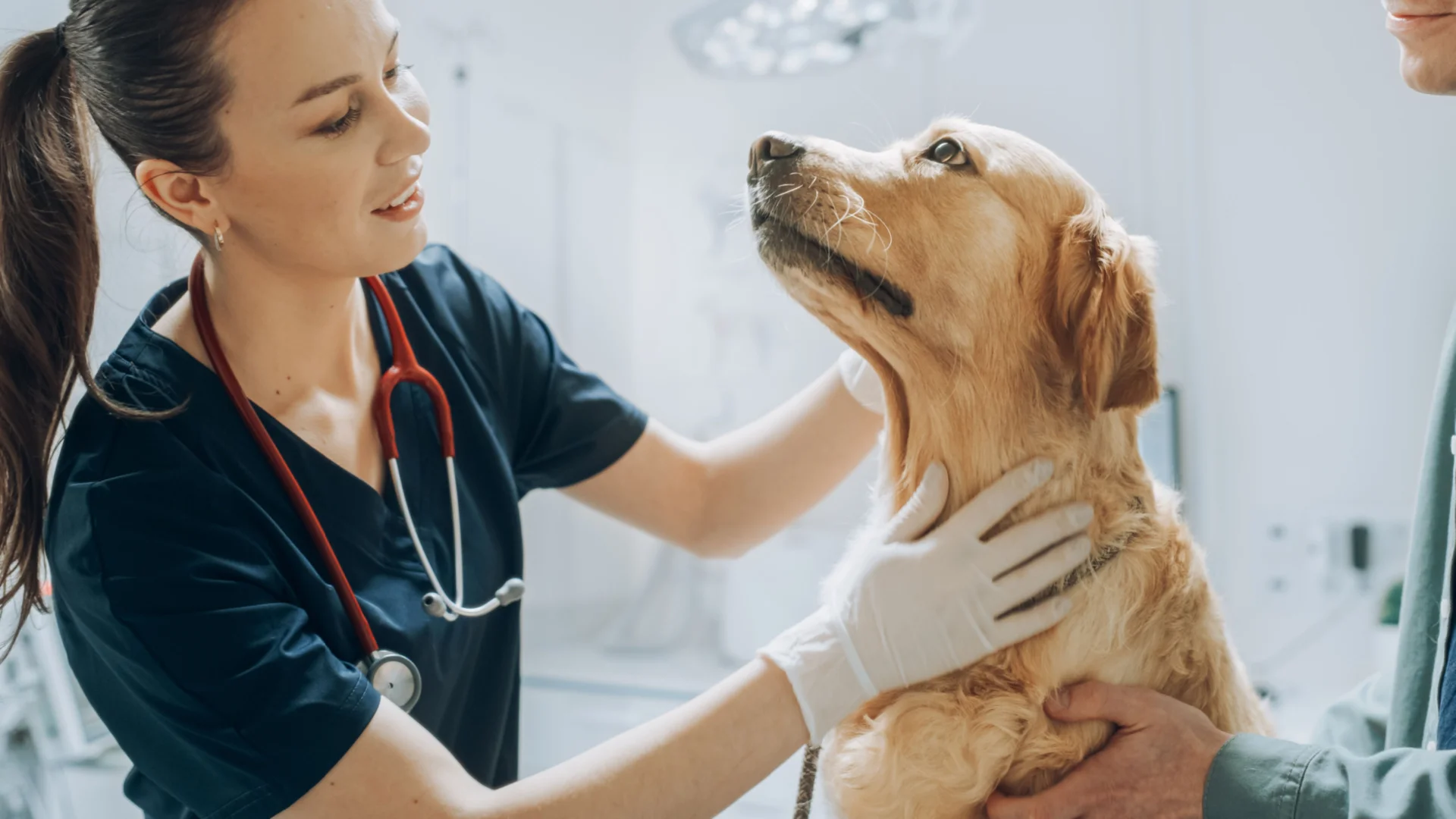
This condition is painful for your scaly friend and if left untreated can result in serious health complications, and in severe cases even death. If you suspect your pet has stomatitis, it is recommended that you make an appointment with your exotic pet veterinarian to diagnose and provide a treatment plan.
What is Stomatitis? (aka Mouth Rot)
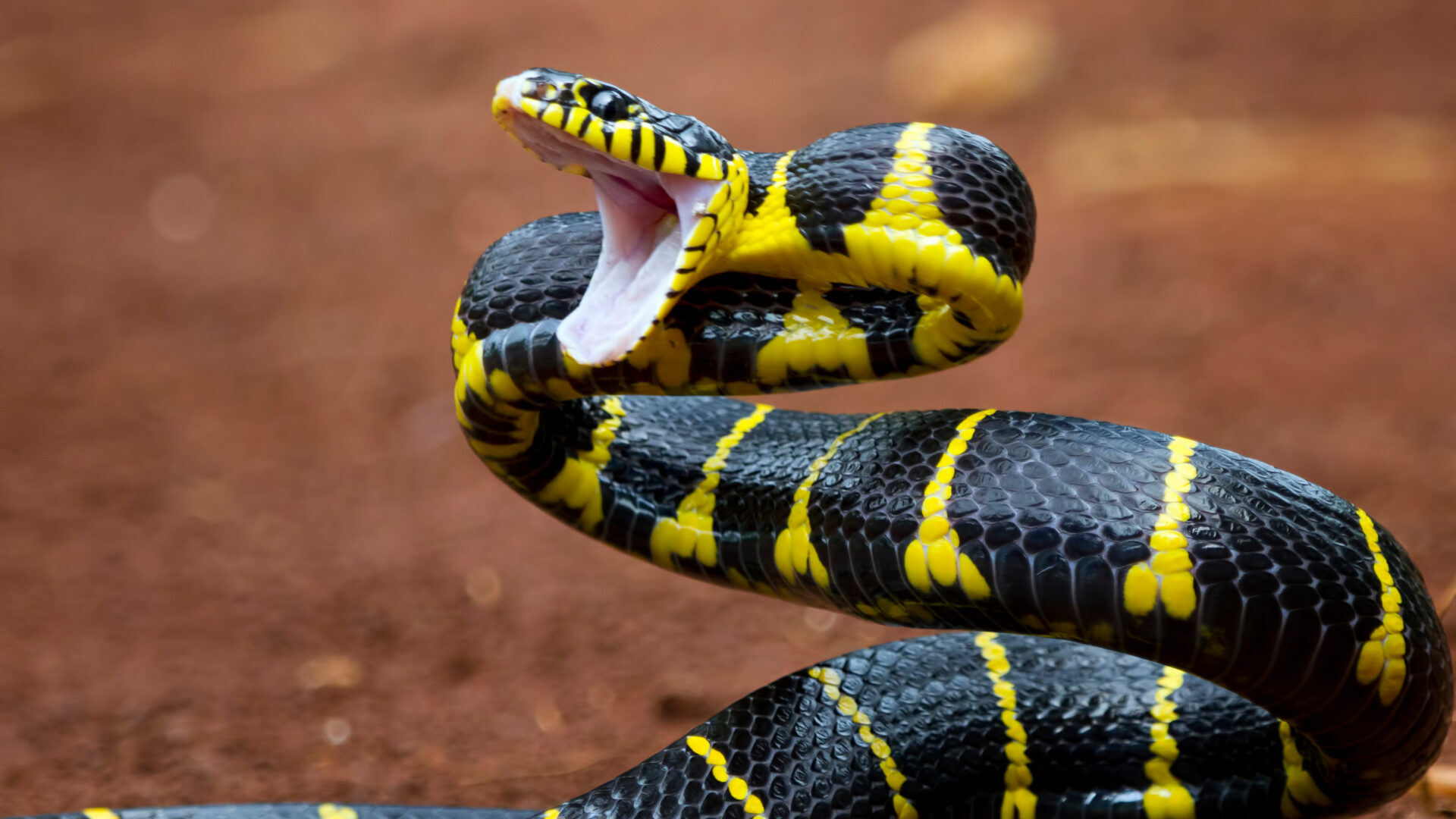
Stomatitis is common in reptiles, most notably afflicting snakes, lizards, and turtles. Mouth rot is mostly caused by opportunistic bacteria infecting reptiles with weakened immune systems, with viruses complicating the severity of infection as well. Specifically, Gram-negative bacilli have been implicated in stomatitis manifestation. Moreover, Aeromonas is among the bacteria implicated as the classical causative agent of infectious stomatitis, along with several other bacteria such as Pseudomonas, Salmonella, Klebsiella, and Mycobacterium [3,4]. As ingested diet and the oral flora have a direct impact on the oral microbiota of your reptile, it is important to monitor your pet’s diet and environmental conditions to encourage a diverse oral microbiome [4]. A review on viruses infecting reptiles found that several viruses have been associated with stomatitis development, and include ranavirus, herpesviruses, adenovirus, papillomaviridae, picornaviridae, and west nile virus [4]. It is important to clarify that more research is needed to determine if these viruses simply leave the reptile more susceptible to opportunistic bacteria, or if there is another mechanism causing the stomatitis infection.
For any reptile, identifying symptoms of stomatitis is critical. Symptoms include:
- Swelling in gums, mouth, and/or nose
- Yellow pus within the mouth (aka cottage cheese mouth)
- Reddened tissue in the mouth
- Increased mucosal drainage from mouth and nose
- Lesions in the oral cavity
- Loss in appetite
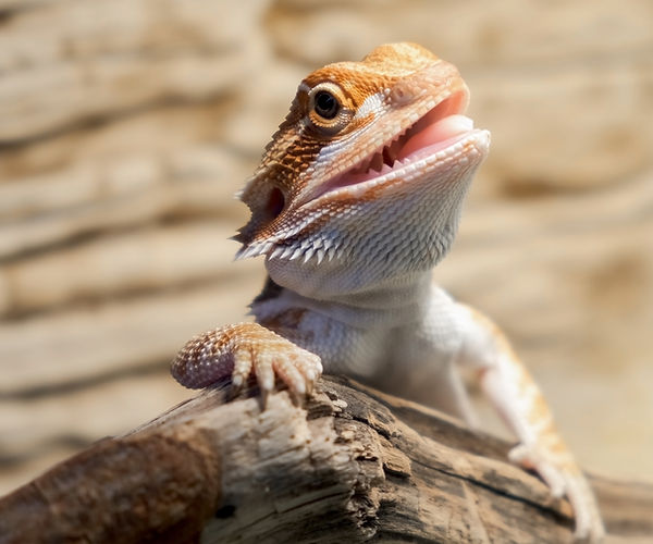
The image above depicts the mouth of a healthy bearded dragon in his terrarium.
Prevention of Stomatitis
Pet reptiles are highly at risk for developing infectious stomatitis due to incorrect temperature and humidity levels. As the Manual of Exotic Pet Practice puts it, “the majority of diseases observed in captive reptiles are directly associated with improper husbandry” [2]. Consequently, setting the proper temperature and humidity levels is key for your pet to maintain a healthy status, as an inability to do so allows for bacteria and fungi to thrive. A proper diet is also very important in maintaining optimal health for your reptile, as vitamin deficiencies can leave your reptile vulnerable to opportunistic pathogens. Oral injuries can also allow openings for pathogenic bacteria to infect your pet, and so monitoring possible ways your reptile is scratching its mouth is useful. And while it may seem obvious that cleaning your pet’s terrarium is important in warding off infections, cleanliness cannot be understated.
Treatment of Stomatitis
Considering the serious nature of stomatitis, it is important to consult with a veterinarian as soon as possible. Delay in care is painful and may result in further medical complications. While treatment will likely entail oral or injectable antibiotics after your pets’ mouth has been cleaned with antiseptics, in severe cases, your reptile may need surgery to remove necrotic tissue. Supportive therapy may be necessary if your reptile is unable to eat or drink properly.
Your veterinarian will also help you identify possible lifestyle changes you and your reptile can make to improve their quality of life and lessen the risk of recurrent infections. This entails understanding the exact pathogen that is impacting your reptile, with modern technological advances allowing for more targeted clinical diagnostic interventions.
Diagnostics
With the rise of antibiotic-resistant strains of pathogens, advances in recent clinical diagnostics have allowed for more targeted and efficient interventions [5]. Historically, culture-based methods have been used to assess the oral microbiome of reptiles, but there are notable diagnostic shortcomings. A recent study assessing serpentovirus (nidovirus) and orthoreovirus coinfection in captive veiled chameleons with the respiratory disease found that “traditional diagnostic assays did not yield evidence of possible contributing infectious causes” [6]. However, metagenomics sequencing allowed the researchers to discover two novel serpentovirus sequences in veiled chameleons [6].
Next-Gen sequencing (NGS) has increasingly helped researchers and veterinarians characterize reptilian oral microbiota [7]. A recent study assessing pneumonia and stomatitis infections in pythons has also affirmed the applicability of NGS technology in characterizing pathogens [8]. This indicates the clinical applicability of using genomic sequencing to identify, analyze, and eventually treat reptiles more effectively. Considering increasing evidence for the efficacy of antimicrobial photodynamic therapy as an alternative treatment for snakes suffering from stomatitis, recognizing the resistance of oral pathogens is extremely useful [5].
Despite its name, the MiDOG All-in-One Microbial Test may provide the answer to the diagnostic conundrum that stomatitis poses. Utilizing NGS technology to detect and quantify all microbial DNA through untargeted and comprehensive sequencing and quantitative comparisons to reference databases, the MiDOG NGS technology provides a useful opportunity to shed light on the microbial makeup of your reptile’s infection for clinical application [9]. The MiDOG microbial test is grounded on scientific research that provides veterinarians DNA evidence for the guided treatment of reptile infections, such as stomatitis.

Stomatitis is difficult to diagnose, but luckily the MiDOG Swab Collection Kit can help!
Find out if your vet uses MiDOG before you book your next appointment!
For health-related questions about your reptile or other exotic pet, reach out to a veterinarian that specializes in exotic pets.
References:
[1] Schmidt V, Klasen L, Schneider J, Hübel J, Pees M. Fungal dermatitis, glossitis and disseminated visceral mycosis caused by different Metarhizium granulomatis genotypes in veiled chameleons (Chamaeleo calyptratus) and first isolation in healthy lizards. Vet Microbiol. 2017 Aug;207:74-82. doi: 10.1016/j.vetmic.2017.06.005. Epub 2017 Jun 10. PMID: 28757044.
[2] Deczm, M. M. D. M. P. & Tully Jr. DVM MS DABVP (Avian) DECZM (Avian), Thomas N. (2008). Manual of Exotic Pet Practice (1st ed.). Saunders.
[3] Singh, J., et. al. (2018). Infectious stomatitis in an Indian rock python (Python molurus) and its therapeutic management. 392-394.
[4] Marschang, R., 2011. Viruses Infecting Reptiles. Viruses, 3(11), pp.2087-2126.
[5] Grego, K., Carvalho, M., Cunha, M., Knöbl, T., Pogliani, F., Catão-Dias, J., Sant’Anna, S., Ribeiro, M. and Sellera, F., 2017. Antimicrobial photodynamic therapy for infectious stomatitis in snakes: Clinical views and microbiological findings. Photodiagnosis and Photodynamic Therapy, 20, pp.196-200.
[6] Sigler, L., Gibas, C. F. C., Kokotovic, B., & Bertelsen, M. F. (2010). Disseminated Mycosis in Veiled Chameleons (Chamaeleo calyptratus) Caused by Chamaeleomyces granulomatis, a New Fungus Related to Paecilomyces viridis. Journal of Clinical Microbiology, 48(9), 3182–3192. https://doi.org/10.1128/jcm.01079-10
[7] Artavia-León, A., Romero-Guerrero, A., Sancho-Blanco, C., Rojas, N. and Umaña-Castro, R., 2017. Diversity of Aerobic Bacteria Isolated from Oral and Cloacal Cavities from Free-Living Snakes Species in Costa Rica Rainforest. International Scholarly Research Notices, 2017, pp.1-9.
[8] Blahak, S., Jenckel, M., Höper, D., Beer, M., Hoffmann, B. and Schlottau, K., 2020. Investigations into the presence of nidoviruses in pythons. Virology Journal, 17(1).
[9] Melgarejo, T., et al., 2021. Canine Urine Microbiome: Assessment of Bacterial and Fungal Populations in Clinically Healthy Dogs Using Next-Generation-Sequencing. Journal of Veterinary Internal Medicine. IN PRESS
Categories: Exotic Pets, Next-Gen DNA Sequencing Technology, Oral/Mouth Infections, Reptiles/Amphibians