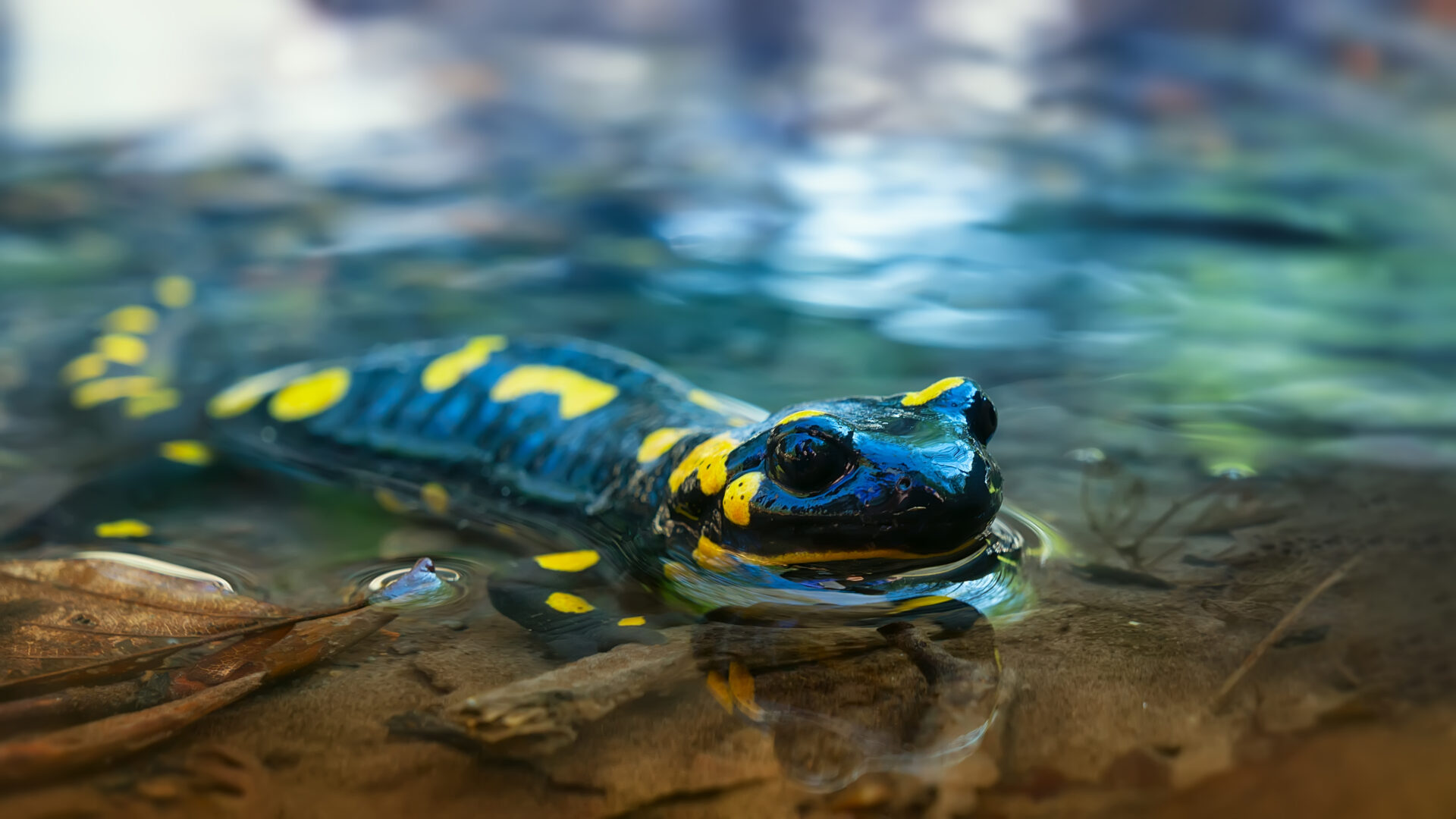
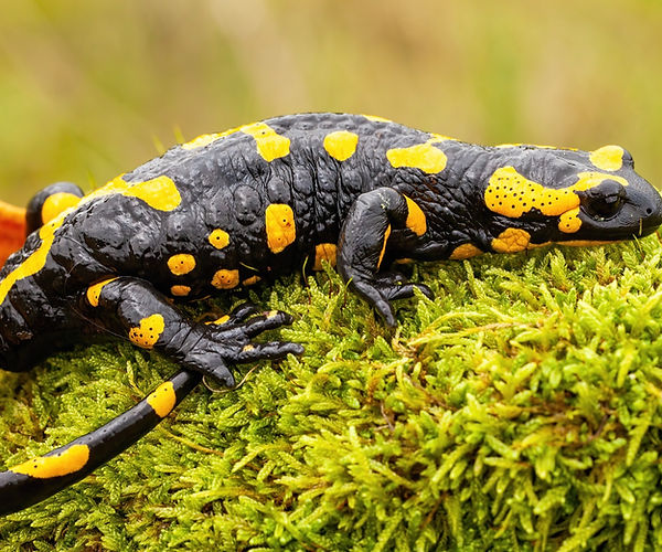
As one of the most common diseases in amphibians, bacterial dermatosepticemia is a fatal infectious disease seen in frogs, toads, and salamanders [1]. More commonly known as red-leg syndrome, bacterial dermatosepticemia is associated with dilation of the capillaries under their skin, which causes the characteristic redness on the underside of the amphibian’s legs and abdomen that is associated with the infectious disease [2]. While the initial exposure to opportunistic bacteria may be caused by physical trauma in the skin, environmental factors such as misguided caretaking and nutritional care create the framework for infectious agents to thrive [3]. Therefore, a combination of understanding the conditions that caused the infection and the infectious pathogens themselves is critical in delivering quality healthcare to your amphibian.
This condition is painful for your amphibian friend and if left untreated can result in serious health complications and death. If you suspect your pet has red-leg syndrome, it is recommended that you make an appointment with your exotic pet veterinarian to diagnose and provide a treatment plan.
What is Bacterial Dermatosepticemia? (aka Red-leg Syndrome)
Bacterial dermatosepticemia is common in amphibians, most notably afflicting frogs, salamanders, and toads. Red-leg syndrome is mostly caused by opportunistic bacteria infecting amphibians with weakened immune systems, with viruses complicating the severity of infection as well. Specifically, Gram-negative bacilli have been implicated in bacterial dermatosepticemia manifestation [4]. Moreover, Aeromonas hydrophila is among the bacteria implicated as the classical causative agent of bacterial dermatosepticemia, along with several other bacteria such as Chryseobacterium indologenes, Chryseobacterium meningosepticum, Citrobacter freundii, Klebsiella pneumoniae, Proteus mirabilis, Pseudomonas aeruginosa, and Serratia liquefaciens [4]. Gram-positive bacteria such as Enterococcus, Streptococcus, and Staphylococcus areus have also been linked to this syndrome [5].
It is important to note that all these bacterial species may be present on the microbiome of your amphibian’s skin or intestine when your pet is healthy; disease progression is often attributable to not only the virulence of the organism but also environmental factors such as stress, diet, health-state, and immune status of your amphibian [2]. For example, A. hydrophila is a common contaminant of water, and so proper animal husbandry is crucial [2].
For any amphibian, identifying symptoms of bacterial dermatosepticemia is critical. The prognosis for this syndrome is poor, with mortality rates ranging from 81% to 100% [6,7]. Therefore, early identification is critical. Notably, acute cases may present more subtle symptoms. Symptoms include:
- Reddening of legs and abdomen
- Hemorrhages in the skeletal muscles, tongue, and nictitating membrane
- Weight loss
- Collection of fluid in the abdominal area
- Open lesions that have not healed
- Loss in appetite
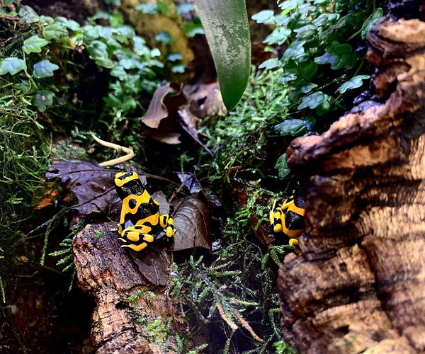
The image above depitcts a black and yellow poison dart frog in a properly maintained vivarium.
How to Prevent Bacterial Dermatosepticemia (Red-leg Syndrome)
Pet amphibians are highly at risk for developing infectious stomatitis due to incorrect temperature and humidity levels. As the Manual of Exotic Pet Practice puts it, “illness in captive amphibians is largely due to poor husbandry practices” [3]. Consequently, setting the proper temperature and humidity levels is key for your pet to maintain a healthy status, as an inability to do so allows for bacteria and fungi to thrive. Amphibians can be aquatic, semiterrestrial, fossorial, terrestrial, or arboreal; therefore it is highly recommended you consult with a veterinarian to get an appropriate understanding of your amphibian’s specific requirements [3].
A proper diet is also very important in maintaining optimal health for your amphibian, as vitamin deficiencies can leave your amphibian vulnerable to opportunistic pathogens. Physical injuries can also allow openings for pathogenic bacteria to infect your pet, and so monitoring possible ways your amphibian is getting scratched is beneficial. And while it may seem obvious that cleaning your pet’s vivarium is important in warding off infections, cleanliness cannot be understated.
Read the Long Island Bird & Exotics Veterinary Clinic Guide here to learn everything you need to know about proper care for your frog or gecko.
Treatment for Bacterial Dermatosepticemia (Red-leg Syndrome)
Considering the serious nature of bacterial dermatosepticemia, it is important to consult with a veterinarian as soon as possible. Delay in care is painful and may result in further medical complications. While treatment will likely entail broad-spectrum antibiotics if a fungal infection is suspected medicated baths may be recommended [5].
Your veterinarian will also help you identify possible lifestyle changes you and your amphibian can make to improve their quality of life and lessen the risk of recurrent infections. This entails understanding the exact pathogen that is impacting your amphibian, with modern technological advances allowing for more targeted clinical diagnostic interventions.
Diagnosing Bacterial Dermatosepticemia (Red-leg Syndrome)
With the clock ticking when attempting to impart a proper diagnosis of bacterial dermatosepticemia, advances in recent clinical diagnostics have allowed for the possibility of more targeted and time-efficient interventions. Historically, culture-based methods have been used to assess the skin and intestinal microbiome of amphibians, but there are notable diagnostic shortcomings. A recent study characterizing the skin microbiome in Italian stream frogs infected and uninfected by a cutaneous parasitic disease confirmed that “next-generation sequencing technologies are advantages [to culture-based approaches] because they provide a thorough description of microbial communities, including uncultivable members” [8]. Specifically, culture-dependent studies reported only 3 different phyla from the frogs studied, while 16S rRNA gene sequencing uncovered 10 to 18 unique skin bacterial phyla, dependent on the species [8].
Next-Gen Sequencing (NGS) has increasingly helped researchers and veterinarians characterize amphibian microbiota. Another study assessing the skin microbiome in a coldspot of amphibian chytriodyccosis infection has also affirmed the applicability of NGS technology in characterizing pathogens [9]. This indicates the clinical applicability of using genomic sequencing to identify, analyze, and eventually treat amphibians more effectively. Considering increasing evidence for highly recombinant pathogen lineages in amphibian populations, recognizing the resistance of bacterial dermatosepticemia-associated bacteria is extremely useful.
Despite its name, the MiDOG All-in-One Microbial Test may provide the answer to the diagnostic conundrum that bacterial dermatosepticemia poses. Utilizing NGS technology to detect and quantify all microbial DNA through untargeted and comprehensive sequencing and quantitative comparisons to reference databases, the MiDOG NGS technology provides a useful opportunity to shed light on the microbial makeup of your amphibian’s infection for clinical application. The MiDOG microbiome test is a microbial identification test grounded on scientific research that provides veterinarians DNA evidence for the guided treatment of amphibian infections, such as bacterial dermatosepticemia.
Find out if your vet uses MiDOG before you book your next appointment!
For health-related questions about your pet, reach out to an exotic pet veterinarian.
References:
[1] Densmore, C., & Green, D. (2007). Diseases of Amphibians. ILAR Journal, 48(3), 235-254. doi: 10.1093/ilar.48.3.235
[2] Whitaker, B., & Wright, K. (2019). Amphibian Medicine. Mader’s Reptile And Amphibian Medicine And Surgery, 992-1013.e3. doi: 10.1016/b978-0-323-48253-0.00089-1
[3] Mitchell, M., & Tully Jr., T. (2009). Manual of Exotic Pet Practice [Ebook] (pp. 78-80). St. Louis: Saunders. Retrieved from https://books.google.com/books?id=8R0_ni8bINAC&pg=PA73&source=gbs_toc_r&cad=3#v=onepage&q&f=false
[4] Schadich, E., & Cole, A. L. (2010). Pathogenicity of Aeromonas hydrophila, Klebsiella pneumoniae, and Proteus mirabilis to brown tree frogs (Litoria ewingii). Comparative medicine, 60(2), 114–117.
[5] Whitaker, B. (2021). Infectious Diseases of Amphibians – Exotic and Laboratory Animals – Veterinary Manual. Retrieved 27 May 2021, from https://www.merckvetmanual.com/exotic-and-laboratory-animals/amphibians/infectious-diseases-of-amphibians
[6] Drake GJ, Koeppel K, Barrows M. Disinfectant (F10SC) nebulization in the treatment of ‘red leg syndrome’ in amphibians. Vet Rec 166(19):593-594, 2010.
[7] Wright KM. Septicemia. In: Mayer J, Donnelly TM (eds). Clinical Veterinary Advisor: Birds and Exotic Pets. St. Louis, MO: Saunders; 2012: 65-67.
[8] Federici, E., Rossi, R., Fidati, L., Paracucchi, R., Scargetta, S., & Montalbani, E. et al. (2015). Characterization of the Skin Microbiota in Italian Stream Frogs (<i>Rana italica</i>) Infected and Uninfected by a Cutaneous Parasitic Disease. Microbes And Environments, 30(3), 262-269. doi: 10.1264/jsme2.me15041
[9] Mutnale, M., Reddy, G., & Vasudevan, K. (2021). Bacterial Community in the Skin Microbiome of Frogs in a Coldspot of Chytridiomycosis Infection. Microbial Ecology. doi: 10.1007/s00248-020-01669-5
Categories: Exotic Pets, Next-Gen DNA Sequencing Technology, Reptiles/Amphibians