MiDOG technology has revolutionized infectious disease diagnostics for exotic pets.
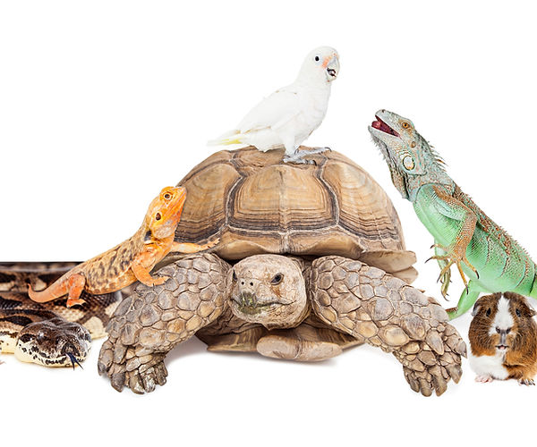
Over the years, exotic animal species have increasingly entered domestic environments as pets. Between 2011 and 2016, there was a reported 26% increase in specialty and exotic pets and a 14% increase in reptilian pets.[1]
This trend highlights the increasing importance of rapid, reliable diagnostic tests to address the corresponding growth of clinical cases involving exotic species. This article aims to provide a brief summary of both cell culture growth and PCR assays as diagnostic tools, their application for exotic and avian clinical cases, and the emergence of Next-Gen DNA sequencing as a robust, superior diagnostic technique.
Cell Culture Testing
Unreliable Growth
Cell culture for clinical diagnosis of infections has persisted for nearly two centuries and currently remains a routine diagnostic tool for the identification of pathogens. While the method is well established in veterinary medicine and can work well for providing insight into the primary pathogenic taxa affecting a patient, it is subject to certain limitations. These limitations can hinder conclusive diagnoses based on cell culture results alone.
The primary limitation of cell culture tests is that they do not always yield results. Many bacteria and fungi are difficult to culture and oftentimes yield ‘no growth’ negative results despite histological confirmation of their presence in tissue samples of an infected area. Mycobacteria, for example, have been reported as slow-growing and often unsuccessfully identified in cell culture tests.[2] Additionally, avian or reptilian patients display body temperatures on either extreme of the norm, making traditional cell culture growth performed at the standard 37°C incubation temperature even more likely to result in no growth for strains adapted to such outlying temperatures.
MiDOG technology is particularly important for exotic pets, who are particularly prone to chronic infections.
Recognizing the increasing trend of exotic pet ownership in the UK, Cushing and colleagues performed a cell culture growth study conducted over 24 months to evaluate microbial diversity in reptiles.[3] Their findings show that out of 254 reptile samples taken for bacterial and fungal growth analysis, 17.6% of fungal cultures and 15.9% of bacterial cultures displayed no growth. The gross failure rate out of the original 254 samples was 18.7%.
Limited Pathogen Detection and Turnaround Time
Other significant limitations of culture testing are the lack of sensitivity to multiple organisms and the variability in the time required to observe growth. While most bacterial cell cultures require 1-3 days to display results, certain strains of bacteria or fungi require up to 5 days to form colonies. Some fungi can even take weeks to grow in laboratory settings.
Aspergillus is one of the most common respiratory mycotic infections observed in birds, and yet in a study conducted on over 300 patients, culture sensitivity was found to be only 40%, with 90% specificity, and a 24.4% positive predictive value.[4] If an initial growth test yields no growth overnight, additional tests using different media, temperature optimization, or anaerobic growth conditions can result in even longer wait times, and may ultimately result in no growth at all.
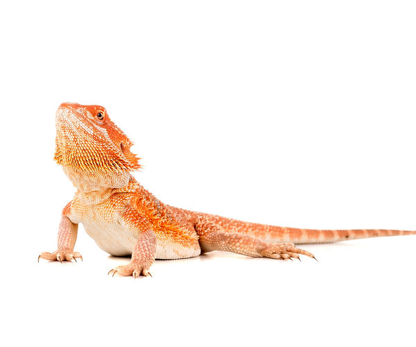
MiDOG technology is superior to culture-based studies, especially when diagnosing exotic animals.
The ability of culture testing to correctly identify all pathogens in an infection is limited, since it has been shown that growth observed from cell cultures might represent as little as 1% of the microbial diversity present in the sample.[5] The lack of sensitivity to multiple pathogenic species means a much greater likelihood of ineffective treatment in patients with a polymicrobial infection. Without knowledge of the entire bacterial and/or fungal population causing the infection, treatment of the predominant pathogen with antibiotics will often only suppress symptoms and allow the strains to develop antibiotic resistance.
PCR as a Diagnostic Tool
Traditional Use and Benefits of PCR as a Diagnostic Procedure
Realizing the above limitations of culture testing, PCR assays have become an increasingly popular diagnostic test for a variety of reasons. Importantly, PCR does not rely on culturing any microbes in the laboratory and detection of pathogens in mixed microbial samples via targeted PCR is extremely sensitive due to the level of DNA amplification that can be achieved over many PCR cycles. As demonstrated by a clinical trial evaluating fecal shedding of Salmonella in horses, PCR sensitivity greatly outweighs culture testing.[6] Unlike serological testing, which relies on the detection of antibodies and can often suffer from a lack of sensitivity and specificity, PCR diagnosis demonstrates an additional advantage by enabling the diagnosis of immunocompromised patients that are unable to produce antibodies.[7,8]
In addition to its versatility and detection power, PCR can also be performed rapidly and quantitatively. Quantitative PCR makes use of fluorescent indicators, such as DNA binding dyes or probes, that allow for real-time detection of the amplified DNA with every cycle. Once a tissue sample or swab of an infected region is submitted for processing, the DNA is amplified and a report sent out within a day or two.
One of the key benefits of using quantitative PCR is the ability to determine the concentration of a pathogen, which can provide insight into the relative timeline of infection and how long the pathogen has proliferated in the host. The rapidity of testing also greatly benefits patients suffering from acute symptoms as other diagnostic methods may simply take too long to yield results before the patient’s condition deteriorates.
Limitations of PCR Diagnosis in Exotic Patients
Despite the advantages of PCR, infections in exotic and avian species can still be difficult to diagnose. The lack of diversity within the targets of common PCR panels used for diagnosis may miss infections in exotic patients. Exotic and avian pets are differentially susceptible to common infections and may be more prone to infection with less common fungal or bacterial pathogens.[9] Since the capacity of PCR panels is limited and cannot contain hundreds of targets, only the most common infectious diseases are screened for, resulting in the underrepresentation of exotic species-related pathogens.
The addition of new pathogen targets to an existing PCR panel is costly. Veterinarians often have to make an educated guess on which pathogens may be causing the infection when selecting the specific pathogen panel for their diagnosis. These panels are often limited to 8 targets, while some offer up to 38 different targets. When the results return with no hits, the game starts over to select a new target panel, with the inherent problem that you will only find what you are looking for. Each additional panel is an added cost for the pet owner, who may be questioning whether the number of tests ordered, and the associated added costs, are truly necessary.
Next-Generation DNA Sequencing in Clinical Diagnostics
Next-Generation DNA sequencing, or Next-Gen DNA sequencing, is a new and robust technique that uses the DNA sequences of every microbe present in a sample to generate a complete picture of the microbial community present. This technique offers the diagnostic specificity and sensitivity of PCR assays without the drawback of limited taxonomic identification and with a similar 2-day turnaround time.
The power of Next-Gen DNA sequencing lies in the taxonomic resolution and determination of relative microbial abundance that is achieved without the need of selecting specific targets. Each conserved DNA sequence identifies the species of the bacteria and fungi from which it originated, and the number of copies of each sequence can be used to determine the relative abundance of each bacteria or fungi within the sample.
Specificity and Sensitivity Without a Limited Panel
Traditional PCR diagnostics target a sequence uniquely present in a certain microbe and demonstrate its presence with PCR amplification. DNA sequencing targets conserved regions of DNA and amplifies the DNA across all microbes. While these regions are very conserved across microbes, they still contain an intrinsic genetic variability that allows each sequence to be assigned to a specific taxon.

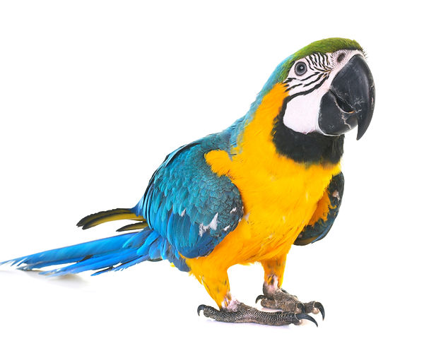
MiDOG technology can help diagnose avian bacterial and fungal infections.
Upon submission of a specimen, such as a skin or cloaca swab, the fungal and bacterial cells present in the sample are mechanically lysed and the extracted DNA is purified. PCR is used to amplify specific regions of DNA (i.e. the 16S ribosomal RNA gene found in all bacteria), and the amplified DNA is then sequenced. The exact sequence of amplified DNA is compared to a comprehensive database of microbial sequences to identify which organisms are present in a sample.
The abundance profile of all microbes within a sample is extremely valuable information in making treatment decisions. Unlike a cell culture growth test, where only one predominant microbe would appear despite the potential presence of dozens of other species, DNA sequencing allows for a complete picture of the abundance of each microbe.
Applicability to Exotic Pets
Based on the untargeted nature of Next-Gen DNA sequencing, it can be applied to any type of clinical sample, from any animal species. The MiDOG All-in-One Test uses precisely this type of technology to analyze clinical samples from exotic animals. The methodology is easily be applied to exotic and avian patients for which traditional diagnostics have been shown to have limited efficacy. This technology offers a solution to the growing number of exotic species-related clinical cases associated with the rising trend of exotic pet ownership. In addition to reporting a ‘detected’ result like PCR panels do, the MiDOG Test adds absolute and relative quantification of all detected microbes, including all potential pathogens. By using this test as a diagnostic tool, you can be positive that all pathogens are identified in your clinical samples.
References:
1. “Section 1.” AVMA Pet Ownership and Demographics Sourcebook: 2017-2018 Edition, AVMA, Veterinary Economics Division, 2018, p. 16.
2. Christine L. Densmore, David Earl Green, Diseases of Amphibians, ILAR Journal, Volume 48, Issue 3, 2007, Pages 235–254, https://doi.org/10.1093/ilar.48.3.235
3. Cushing, A., et al. “Review of Bacterial and Fungal Culture and Sensitivity Results from Reptilian Samples Submitted to a UK Laboratory.” Veterinary Record, vol. 169, no. 15, 2011, pp. 390–390., doi:10.1136/vr.d4636.
4. Levy, H., et al. “The Value of Bronchoalveolar Lavage and Bronchial Washings in the Diagnosis of Invasive Pulmonary Aspergillosis.” Respiratory Medicine, vol. 86, no. 3, 1992, pp. 243–248., doi:10.1016/s0954-6111(06)80062-4.
5. Barcina, Isabel, and Inés Arana. “The Viable but Nonculturable Phenotype: a Crossroads in the Life-Cycle of Non-Differentiating Bacteria?” Reviews in Environmental Science and Bio/Technology, vol. 8, no. 3, 2009, pp. 245–255., doi:10.1007/s11157-009-9159-x.
6. Ward, Michael P., et al. “Evaluation of a PCR to Detect Salmonella in Fecal Samples of Horses Admitted to a Veterinary Teaching Hospital.” Journal of Veterinary Diagnostic Investigation, vol. 17, no. 2, 2005, pp. 118–123., doi:10.1177/104063870501700204.
7. Díaz-Aparicio, E, et al. “Evaluation of Serological Tests for Diagnosis of Brucella Melitensis Infection of Goats.” Journal of Clinical Microbiology, vol. 32, no. 5, 1994, pp. 1159–1165., doi:10.1128/jcm.32.5.1159-1165.1994.
8. Pena, Hilda Fátima De Jesus, et al. “Fatal Toxoplasmosis in an Immunosuppressed Domestic Cat from Brazil Caused by Toxoplasma Gondii Clonal Type I.” Revista Brasileira De Parasitologia Veterinária, vol. 26, no. 2, 2017, pp. 177–184., doi:10.1590/s1984-29612017025.
9. Troxell, Bryan, et al. “Poultry Body Temperature Contributes to Invasion Control through Reduced Expression of Salmonella Pathogenicity Island 1 Genes in Salmonella Enterica Serovars Typhimurium and Enteritidis.” Applied and Environmental Microbiology, vol. 81, no. 23, 2015, pp. 8192–8201., doi:10.1128/aem.02622-15.
Categories: Birds/Parrots, Exotic Pets, Reptiles/Amphibians, Veterinarian Guides, Veterinary Best Practices
