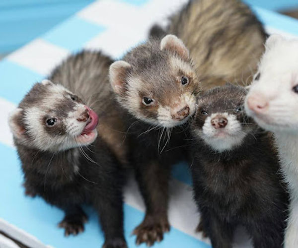
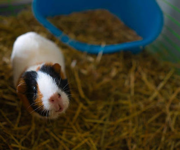
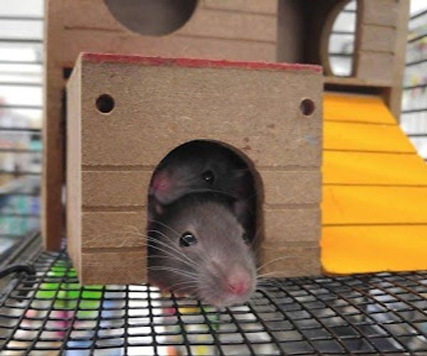
Bumblefoot (pododermatitis) is a skin infection on the feet of small pets like guinea pigs, rats, hamsters, ferrets, rabbits, and chinchillas. Here’s what you need to know about treating bumblefoot effectively and confidently in small exotic pets.
What are the symptoms of bumblefoot in small exotic animals?
In the early stages of bumblefoot, noticeable signs include swelling and redness on the footpad. A characteristic feature is the emergence of a swollen, pus-filled bump, which is often accompanied by a central, dark-colored scab. Animals experiencing lameness or putting more weight on a given leg(s) are more susceptible to bumblefoot infections.
Left untreated, the condition progresses to a late stage, posing severe risks to the musculature and bones in the foot. Tissues and tendons can become weakened or necrotic as the infection spreads toward the bone (osteomyelitis). The bone infection can cause sepsis and death. [1]
What causes bumblefoot in small exotic animals?
Bumblefoot is typically a secondary infection to an underlying injury. Pathogens can enter the wound and proliferate more easily when the skin is vulnerable. By taking advantage of the skin’s weakness, the pathogen is acting opportunistically.
Such circumstances that allow for opportunistic infections include:
Bite wounds
Male-to-male and male-to-female aggression is common during breeding seasons but can occur at other times of the year. Competition is a means of establishing social hierarchy and dominance, which can result in bite and scratch wounds that can become infected.
Obesity
Excessive weight on foot pads can lead to skin irritation.
Rough surfaces
Standing on solid and rough surfaces (e.g., rough carpet, solid hard floor, and bare wire floor) can cause the skin on the bottom of the feet to become dry, cracked, and blistered. With this added strain, blisters can rupture, allowing for pathogenic organisms to invade the wound.
Long nails
Leaving nails untrimmed can cause uneven weight distribution and cuts on the feet.
Improper bedding
Bedding that doesn’t properly soak up urine, water, and feces increases the risk of infection. Urine, water, and feces host a lot of potentially harmful bacteria that can enter the exposed skin, such as Escherichia coli.
Treatment options for bumblefoot in rodents and small exotic animals
Bumblefoot is commonly a secondary infection resulting from preexisting issues in the animal’s environment or husbandry. The administration of treatment alone will not cure the underlying cause. Addressing the root of infection is crucial for long-term resolution.
A veterinarian should assess the extent of the infection and provide recommendations for treatment both in the clinic and at home. It is imperative to choose all at-home remedies under the guidance from a veterinarian. Here are a few examples of such remedies:
- Warm foot baths with or without Epsom salt.
- Boost Vitamin A intake for skin recovery and strength.
- Boost Vitamin C intake to help fight the infection.
- Utilize a thick moisturizer when the skin has healed to prevent skin dryness and cracking.
- For chinchillas, pause the use of dust baths to curb irritation and dryness in the feet.
Treatment for bumblefoot varies depending on the severity of the infection but typically consists of a combination of topical antimicrobials, surgical drainage, surgical removal, and antibiotics. Choosing the right antibiotic is critical for the patient’s overall health. Altering the normal microbiome can have adverse effects, such as the development of drug-resistant strains and even death. [2, 3]
The challenge with treating bumblefoot with antibiotics arises from the non-specific nature of the infection. Many microorganisms can become problematic when provided with the opportunity to become pathogenic and dominate the microbiome. Staphylococcus aureus [2] and Pasturella multocida [4] commonly cause bumblefoot in small exotic animals, but several other microorganisms are known to cause bumblefoot in other pets.
In order to choose the most effective antibiotic, the bacteria causing the infection must be found first. However, there are some challenges to conventional identification and antibiotic susceptibility testing:
Bacteria have different growth requirements.
It takes time to identify non-specific infections by culture. Even under optimal conditions, culture fails to grow 15% of bacteria and 81% of fungi compared to Next-Generation Sequencing (NGS) [2]. This delay allows for the infection to progress and cause more damage.
Bacteria can acquire antimicrobial resistance genes.
Antimicrobial resistance can be intrinsic to the microorganisms or acquired. Acquired resistance can arise from (1) horizontal gene transfers of resistance genes from foreign bacteria or (2) the mutation of their own genes [3, 5].
These acquired genes are not present in the entire bacterial population and could be missed during culture sensitivity testing. Testing for these genes directly using DNA sequencing can ensure accurate reporting of antibiotic susceptibility.
Cell counts are necessary when deciding which microorganism to target with antibiotics.
Knowing the cell count of each microorganism provides crucial context on which one to target. Quantification is limited in culture testing and not possible with PCR panels.
Fortunately, there is a diagnostic tool capable of addressing these obstacles to identifying pathogens and determining the right treatment option. Microbial DNA sequencing with the MiDOG All-in-One Test allows for detection and quantification of all bacterial and fungal pathogens. No guesswork is needed for the antibiotic profiling of the species of interest either. The MiDOG All-in-One Test includes antibiotic resistance profiling on 43 antibiotics, which informs the microbes’ antibiotic susceptibility.
If you think your patient may be suffering from bumblefoot, consider using the MiDOG test to determine confidently and quickly.
References
[1] Blair, J. (2013). Bumblefoot: A comparison of clinical presentation and treatment of pododermatitis in rabbits, rodents, and birds. Veterinary Clinics of North America: Exotic Animal Practice, 16(3), p. 715-735. https://doi.org/10.1016/j.cvex.2013.05.002.
[2] Scott, D. et.al. (2001). Dermatoses of pet rodents, rabbits, and ferrets. Muller & Kirk’s Small Animal Dermatology, 1415–1458. https://doi.org/10.1016/B978-0-7216-7618-0.50025-0.
[3] Palma, E. et.al. (2020). Antimicrobial resistance in veterinary medicine: An overview. International Journal of Molecular Sciences, 21(6), 1914. https://doi.org/10.3390/ijms21061914.
[4] Schoemaker, N. & Van Zeeland, Y. (2016). Potodermatitis in birds and small mammals. 8th World Congress of Veterinary Dermatology and the World Association for Veterinary Dermatology. Accessed 19 Jan 2024. wcvd8-proceedings.pdf (wavd.org)
[5] Harbottle, H. et.al. (2007). Genetics of antimicrobial resistance. Animal biotechnology, 17(2), 111-124. https://doi.org/10.1080/10495390600957092.