
Even though your dog may not be showing signs, if they have been exposed to unsanitary water or other infected animals, they may be suffering from leptospirosis. Dogs are particularly susceptible to this bacterial disease, which has seen an increasing prevalence with approximately 8.2% of dogs shedding the infectious pathogen [1]. Transmission of leptospirosis is largely due to environmental factors such as high precipitation rates, flooding, uncontrolled urban development, and poor sanitary conditions [1]. Leptospirosis is caused by water-borne bacteria, and so exposure to puddles and moist soil contaminated by the urine of infected animals can cause disease transmission.
This condition is painful for your furry friend and can develop into serious life-threatening conditions affecting the kidneys, liver, heart, lungs, and brain if left untreated [2]. Early intervention is recommended, and so if you suspect your pet has leptospirosis, visit a veterinarian immediately so they can diagnose and provide a treatment plan.
What is Leptospirosis?
Leptospirosis is caused by the pathogenic Leptospira genus, which has been reported to infect over 150 mammalian species and has over 250 serotypes [3]. Leptospira is a flexible, spiral-shaped, Gram-negative bacteria that is extremely pathogenic in canines; specifically, L. interrogans and L. kirschneri infections are the primary culprits in canine infections [4].
Transmission of leptospires occurs through direct contact or indirect contact of the pathogen due to environmental exposures. The pathway that the leptospires take to infect their host is complex, as the pathogen enters the body through mucous membranes in the eyes, nose, mouth, or in some cases skin [1]. The pathogen is then able to multiply in the bloodstream and go on to infect the kidneys and other tissue. The infectious cycle renews once the infected dog sheds the pathogen through urination, potentially exposing other animals to the disease [1]. Notably, some infected dogs may enter a carrier state and be asymptomatic [5]. In some cases, Leptospira spp. are able to persist and shed in urine for weeks to months [1].
Climate change has substantially impacted the transmissibility of the disease by creating weather conditions that allow perfect situations (like floods) for exposure to unsanitary water [6]. Increasing rates of this disease are significantly noted in the Midwest, Eastern, and Southwestern USA [6]. Regardless, dogs with access to ponds, lakes, streams, or other stagnant water are particularly at risk for developing leptospirosis [1]. If left untreated, leptospirosis can result in leptospiremia, which is when leptospires multiply in the bloodstream and go on to infect other tissues and organs. Complications that can result from leptospiremia include vascular damage (and consequently kidney failure, liver damage, and/or clotting problems) and/or acute renal failure and hepatic dysfunction [5]. For any dog parent, identifying symptoms of leptospirosis is critical. Early symptoms include:
- Fever
- Lethargy
- Anorexia/weight loss
- Depression
- Acute renal failure
- Jaundice
- Abdominal discomfort
- Vomiting and diarrhea
- Respiratory distress
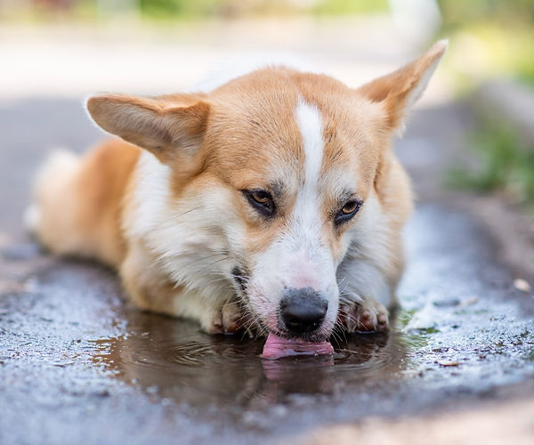
The image above depicts a corgi drinking water from a puddle. To avoid infectious diseases like leptospirosis, it is recommended to avoid drinking from stagnant water.
Preventing and Treating Leptospirosis in Your Dog
Dogs are highly at risk for developing leptospirosis due to exposure to unsanitary conditions. While it may seem harmless to let your dog drink out of stagnant puddles of water on a walk, jump into a pond on a hike, etc., this can lead to unnecessary exposure to Leptospira spp. It is especially important to be aware of possible exposures to the pathogen, as a treatment for leptospirosis is admittedly lacking.
Early diagnosis allows for the administration of procaine benzylpenicillin G, which over weeks may cure the infection and help curb organ damage. Should liver damage occur, further treatment is needed [5]. Generally, doxycycline is the standard treatment for leptospirosis, although curing the infection is difficult. Often times an infected dog may become a chronic carrier of the disease and require long-term monitoring [5].
Diagnosing Leptospirosis in Your Dog
Historically, culture-based methods have been used to assess possible bacterial infections in dogs, but there are notable diagnostic shortcomings. In a prospective study of canine leptospirosis using both culture and PCR techniques, the authors noted how “culturing of leptospires, albeit essential to confirm infection, is not a suitable technique for the identification of urinary shedders, especially for presenting frequent contamination, low sensitivity, and fastidious growth of the pathogen” [7]. PCR testing also has severe shortcomings in terms of leptospirosis diagnostics, as intermittent shedding may lead to false-negative PCR results [7]. Conversely, Next-Gen Sequencing (NGS) has provided an exciting and effective alternative to culture- and PCR- based diagnostics, allowing for improved Leptospira classification and diagnostics [8].
The MiDOG All-in-One Microbial Test may provide the answer to the diagnostic conundrum that leptospirosis poses for your dog. Utilizing NGS technology to detect and quantify all microbial DNA through untargeted and comprehensive sequencing and quantitative comparisons to reference databases, the MiDOG NGS technology provides a useful opportunity to shed light on the microbial makeup of your dog’s bacterial infection for clinical application. The MiDOG microbial test is grounded on scientific research that provides veterinarians DNA evidence for the guided treatment of dog infections, such as leptospirosis.

Find out if your vet uses MiDOG before you book your next appointment!
References
[1] Canine Leptospirosis | Merck Animal Health USA. Merck Animal Health USA. (2021). Retrieved from https://www.merck-animal-health-usa.com/nobivac/canine-leptospirosis.
[2] Murphy, K. (2018). Leptospirosis in dogs and cats: new challenges from an old bacteria. In Practice, 40(6), 218-229. https://doi.org/10.1136/inp.k2926
[3] Sykes, J. E., Hartmann, K., Lunn, K. F., Moore, G. E., Stoddard, R. A., & Goldstein, R. E. (2011). 2010 ACVIM small animal consensus statement on leptospirosis: diagnosis, epidemiology, treatment, and prevention. Journal of veterinary internal medicine, 25(1), 1–13. https://doi.org/10.1111/j.1939-1676.2010.0654.x
[4] Major, A., Schweighauser, A., & Francey, T. (2014). Increasing Incidence of Canine Leptospirosis in Switzerland. International Journal Of Environmental Research And Public Health, 11(7), 7242-7260. https://doi.org/10.3390/ijerph110707242
[5] Gutierrez, L., Mendoza, J., Rangel, A., Tapia, G., Bernad, M., & Sumano, H. (2019). Outpatient Clinical Trial in Dogs With Leptospirosis Treated With Enrofloxacin Hydrochloride-Dihydrate (ENRO-C). Frontiers In Veterinary Science, 6. https://doi.org/10.3389/fvets.2019.00360
[6] White, A., Zambrana-Torrelio, C., Allen, T., Rostal, M., Wright, A., & Ball, E. et al. (2017). Hotspots of canine leptospirosis in the United States of America. The Veterinary Journal, 222, 29-35. https://doi.org/10.1016/j.tvjl.2017.02.009
[7] Miotto, B. A., Guilloux, A., Tozzi, B. F., Moreno, L. Z., da Hora, A. S., Dias, R. A., Heinemann, M. B., Moreno, A. M., Filho, A., Lilenbaum, W., & Hagiwara, M. K. (2018). Prospective study of canine leptospirosis in shelter and stray dog populations: Identification of chronic carriers and different Leptospira species infecting dogs. PloS one, 13(7), e0200384. https://doi.org/10.1371/journal.pone.0200384
[8] Agampodi, S., & Vinetz, J. (2021). Next-Generation Sequencing Analysis of Pathogenic Leptospira: A Way Forward for Understanding Infectious Disease Dynamics in Low/Middle-Income, Disease-Endemic Settings. The American Journal Of Tropical Medicine And Hygiene, 104(5), 1625-1627. https://doi.org/10.4269/ajtmh.20-1518