Bulldogs, boxers, and pugs, oh my! These are just a few of the many breeds of brachycephalic dogs, defined as dogs with flattened faces [1]. As cute as their smooshed faces may be, these breeds are particularly prone to skin infections (like intertrigo) due to moisture-retaining skin folds that provide the ideal breeding ground for opportunistic bacteria and fungi [1].
A recent study using Next-Gen DNA Sequencing (NGS) was conducted to assess the skin fold microbiome in healthy French bulldogs and determine how the microbiome is influenced by topical medications. Considering the need for alternative approaches to normalizing the epidermal barrier and lessening the severity of intertrigo presentation due to increasingly resistant strains of bacteria, this research holds significant implications for how intertrigo can be diagnosed and treated [2]. Read more to learn about intertrigo and advancements in clinical diagnostics for your wrinkly pup!
So What is Intertrigo?
Intertrigo, also known as skin fold dermatitis, is a common skin condition in brachycephalic dogs caused by a unique combination of epidermal trauma (associated with skin friction) and environmental moisture that allows microbial organisms to thrive [3]. The clinical manifestation of this condition results from an imbalance in the dog’s microbiome, which includes fungi, bacteria, viruses, protozoa, and phages [2]. Notably, a healthy nasomaxillary microbiome can actually prevent the colonization of pathogenic organisms [2]. Intertrigo causes recurrent/continuous episodes of scratching, rubbing, chewing, and licking, ultimately resulting in the inflammation and damage of your dog’s skin [4]. While studies of the nasomaxillary microbiome are limited, current research suggests Staphylococcus, Streptococcus, Pseudomonas, and Malassezia species are implicated in intertrigo manifestation.
Intertrigo can range from being mild to extremely painful, and so it is important to be on the lookout for symptoms that include but are not limited to:
- Excessive itching
- Redness and inflammation
- Irritation in armpits, groin, eyes, ears, snout, and between toes
- Skin lesions, blisters, and/or hives
- Skin and ear infection
- Flaking or crusting skin
If you think your dog may be suffering from intertrigo, please consult with a veterinarian as soon as possible. In some cases, surgical intervention may be necessary to remove excess skin folds that create the environment for intertrigo-associated microbiota to proliferate [5]. A topical antibiotic ointment such as mupirocin may be recommended in cases of severe bacterial overgrowth, but increasing mupirocin resistance in canine staphylococci is raising alarm for treatment interventions [2].
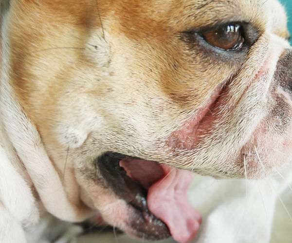
The image depicts inflammation and redness associated with intertrigo.
Characterizing the Nasomaxillary Fold Microbiome
Conventional culture-based methods have been found to have significant limitations in determining the nasomaxillary microbiome, largely due to the variability in culturing different bacteria from a plethora of diverse genera [6]. Moreover, characterizing the fungal organisms in your dog’s skin folds are difficult through culture based methods, as this process not only takes weeks but also can result in no growth results [6]. Recent advances in next-generation sequencing (NGS) technologies allow metagenomics analysis, and have provided exciting and practical opportunities for canine intertrigo interventions.
“Multi-drug antibiotic resistance has further complicated [treatment] and made both topical therapy selection and diagnostic testing of the utmost importance.”
Particularly, a recent study was conducted using MiDOG NGS technology to assess the skin fold microbiome in healthy French bulldogs. This study found that the primary skin bacterial phyla populating the nasomaxillary skin fold were Firmicutes, and Proteobacteria, while the primary skin fungal phyla were Ascomycota and Basidiomycota [2]. Also, the researchers noted a significant correlation between the abundance of potentially opportunistic pathogens and microbial diversity. Healthy French bulldogs contained high ratios of clinically relevant pathogens (36.4%) in their nasomaxillary skin fold microbiome [2].
Additionally, the study looked at how topical treatment impacts the diversity of bacterial and fungal compositions over time, which is clinically useful because it can provide an alternative to antibiotic treatment. The dermal treatments analyzed in this study looked at a protease product (Kalzyme) that inhibits biofilm formation but has no biocidal activity and a 2% chlorhexidine diacetate (Nolvasan; CHX) solution, which has broad spectrum biocidal activity against bacteria and fungi [2]. Interestingly, diversity increased by 38% for the protease group, as opposed to 11% for the CHX group.
Since the topical therapy with protease increases microbial diversity in the skin folds and reduces the relative abundance of pathogens, this therapy could hold clinical value in the treatment of intertrigo. As one of the researchers in this study, veterinary Dr. Alissa Rexo notes how “superficial bacterial pyoderma is one of the most common dermatologic diseases in dogs. Multi-drug antibiotic resistance has further complicated this problem and made both topical therapy selection and diagnostic testing of the utmost importance.” Considering intertrigo is one of the most common forms of surface pyoderma, it is important to ask your veterinarian for more information on treatment options.

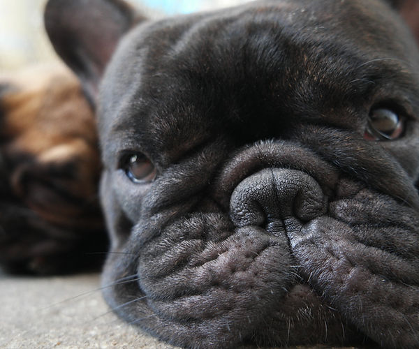
The image above depicts one of the study participants on Day 0 exhibiting the classic nasomaxillary folds that predispose dogs to intertrigo manifestation.
If you would like to read the full study, please follow this link: https://onlinelibrary.wiley.com/doi/abs/10.1111/vde.13017
Moreover, MiDOG NGS technology was also used to analyze the bacterial and fungal microbiota in health and diseased skin in one of the largest canine cohort studies to date. This study found that important taxa enriched in disease-state skin included Malassezia pachydermatis, Staphylococcus pseudintermedius, Staphylococcus schleiferi, and anaerobic bacteria such as Finegoldia magna, Peptostreptococcus canis, and Porphyromonas cangingivalis [7]. These findings are particularly notable, as the anaerobic microorganisms identified to be associated with canine skin infections have previously been overlooked by culture-based studies. The potential of NGS-based methods for the accurate quantification and identification of bacterial and fungal populations in diagnosing canine skin infections is remarkable, and highlight the limitations of traditional culture-based testing.
If you would like to read the full study, please follow this link: https://www.sciencedirect.com/science/article/pii/S0378113519314932
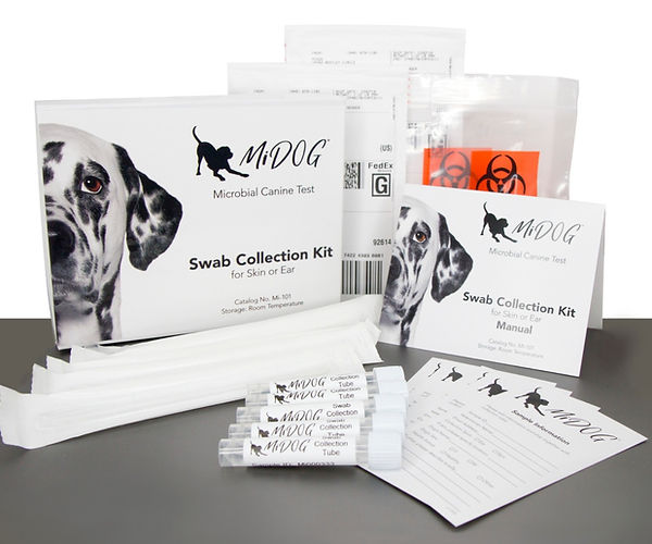
The MiDOG All-in-One microbiome test utilizes the NGS technology as described in the study to detect and quantify all microbial DNA through untargeted and comprehensive sequencing and quantitative comparisons to reference databases. Considering shortcomings in culture-based diagnostics, the MiDOG NGS technology provides a useful opportunity to shed light on the microbial makeup of your dog’s nasomaxillary microbiome for clinical application. The MiDOG microbiome test is a microbial identification test grounded on scientific research that provides veterinarians DNA evidence for the guided treatment of canine infections, such as intertrigo.
Find out if your vet uses MiDOG before you book your next appointment!
References:
[1] Fawcett, A., Barrs, V., Awad, M., Child, G., Brunel, L., Mooney, E., Martinez-Taboada, F., McDonald, B. and McGreevy, P., 2018. Consequences and Management of Canine Brachycephaly in Veterinary Practice: Perspectives from Australian Veterinarians and Veterinary Specialists. Animals, 9(1), p.3.
[2] Rexo, A., Hansen, B., Clarsund, M., Krumbeck, J., & Bernstein, J. (2021). Effect of topical medication on the nasomaxillary skin‐fold microbiome in French bulldogs. Veterinary Dermatology. https://doi.org/10.1111/vde.13017
[3] Gross, T., Ihrke, P., Walder, E., et al., 2005. Skin Diseases of the Dog and Cat. 2nd ed. Oxford: Blackwell Science Ltd; pp261-263.
[4] Anturaniemi, J., Uusitalo, L. and Hielm-Björkman, A., 2017. Environmental and phenotype-related risk factors for owner-reported allergic/atopic skin symptoms and for canine atopic dermatitis verified by veterinarian in a Finnish dog population. PLOS ONE, 12(6), p.e0178771.
[5] Miller, W., Griffen, C., Campbell, K., 2016. Muller and Kirk’s Small Animal Dermatology. 7th ed. St. Loius, MO: Elsevier; pp678-680.
[6] Harvey, N., Shaw, S., Blott, S., Vàzquez-Diosdado, J. and England, G., 2019. Development and validation of a new standardised data collection tool to aid in the diagnosis of canine skin allergies. Scientific Reports, 9(1).
[7] Tang, S., Prem, A., Tjokrosurjo, J., Sary, M., Van Bel, M., Rodrigues-Hoffmann, A., Kavanagh, M., Wu, G., Van Eden, M. and Krumbeck, J., 2020. The canine skin and ear microbiome: A comprehensive survey of pathogens implicated in canine skin and ear infections using a novel next-generation-sequencing-based assay. Veterinary Microbiology, 247, p.108764.