When it comes to treating infectious diseases in marine mammals, like sea lions, traditional diagnostic methods can fall short, particularly when dealing with complex infections like abscesses. These lesions are often polymicrobial, involving a combination of bacterial and fungal microorganisms, many of which are fastidious or unculturable using standard laboratory techniques.
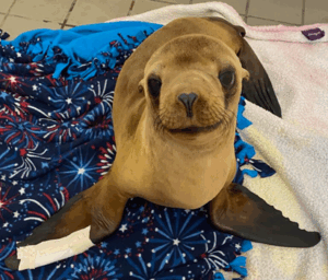
This challenge was exemplified in the case of Hai-Xi, a stranded sea lion pup rescued by the Pacific Marine Mammal Center (PMMC). He arrived malnourished and dehydrated, with a severe, worsening abscess in his right flipper. Despite initial supportive care of rehydration and nutritional support, his condition worsened, and his right flipper continued to deteriorate, prompting the team to seek a more advanced diagnostic solution beyond the initial radiography and blood work.
Why Abscesses in Sea Lions Are Challenging to Diagnose
Abscesses in sea lions may result from trauma, systemic infections, or immunosuppression. Constant exposure to seawater, marine debris, and pathogens in their environment increases the risk of infection from opportunistic or novel microorganisms. Many of these organisms originate from the host’s own microbiota or marine environment.
Traditional culture-based diagnostics often fail to detect the full scope of infection. In fact, fewer than 20–30% of the actual microbial diversity in a marine abscess is typically detected using culture, due in part to:
- Strict anaerobes requiring oxygen-free conditions to grow
- Slow-growing or host-adapted organisms that don’t survive transport or lab conditions
- Fungi that aren’t targeted by standard bacterial cultures and may take weeks to grow, if at all
In Hai-Xi’s case, routine diagnostics weren’t enough to explain the worsening infection and tissue damage. That’s when a sample of his flipper abscess was sent to MiDOG Animal Diagnostics for culture-independent testing.
Common and Uncommon Culprits: Bacteria and Fungi in Marine Abscesses
Thanks to Next-Generation Sequencing (NGS), we now have a clearer picture of the microbial landscape in marine mammal infections. MiDOG Animal Diagnostics uses DNA sequencing to profile both bacterial and fungal communities directly from clinical samples without the need for culturing.
Common bacterial organisms identified in sea lion abscesses using NGS include:
- Fusobacterium nucleatum: A strict anaerobe linked to abscess formation across species.
- Streptococcus phocae: A facultative anaerobe that is commonly seen in marine mammals, like sea lions, and is associated with abscess formation and pneumonia, among other systemic infections
- Clostridium perfringens: A toxin-producing anaerobe often missed in routine diagnostics.
- Bacteroides fragilis: A fastidious anaerobe involved in polymicrobial infections.
- Escherichia coli: An enteric organism that may translocate to extraintestinal sites and contribute to abscess development.
- Aeromonas: Waterborne bacteria capable of causing soft tissue and systemic infections in marine species.
Fungal organisms often found in abscesses include:
- Candida tropicalis: A yeast species that can translocate from mucosal sites and cause localized or systemic infections.
- Debaryomyces hansenii: A halotolerant yeast naturally found in marine environments, now increasingly implicated in soft tissue infections.
- Cladosporium and Aspergillus species: Environmental molds capable of colonizing wounds.
- Malassezia: A lipophilic yeast that is typically part of the skin microbiota but may proliferate in inflamed or disrupted tissues, contributing to chronic infection.
- Mycoplasma: A fastidious bacterium that lacks a cell wall and is often associated with mucosal surfaces and chronic infections; difficult to culture but detectable by molecular methods.
When Hai-Xi’s MiDOG results came back, they revealed high concentrations of Streptococcus phocae– nearly 100% of their skin microbiome! S. phocae is a bacterium that conventional diagnostics often misses or misinterprets at the species level. While S. phocae is technically culturable, it is often misidentified as other β-hemolytic streptococci. The accuracy of NGS testing allowed the clinical team to make targeted decisions about Hai-Xi’s treatment.
The MiDOG Advantage: Culture-Independent Marine Mammal Diagnostics
At MiDOG Animal Diagnostics, we specialize in microbial detection that goes beyond what culture can see. Our NGS-based test captures the full spectrum of bacterial and fungal DNA, enabling veterinarians to identify:
- Unculturable and hard-to-grow microbes
- Anaerobes and low-abundance pathogens
- Polymicrobial interactions relevant to disease progression
- Fungi often missed in routine panels
In Hai-Xi’s case, the identification of Streptococcus phocae via MiDOG testing led to a surgical intervention (amputation of two thumb bones) followed by continued antimicrobial management. A follow-up MiDOG test confirmed that S. phocae was no longer present. His flipper healed, the infection resolved, and Hai-Xi’s condition steadily improved!
Conclusion: Seeing the Full Picture Beneath the Surface
Abscesses in sea lions are often driven by diverse microbial communities that remain hidden to culture-based tests. MiDOG Animal Diagnostics provides a powerful alternative: Next-Generation Sequencing that uncovers the full bacterial and fungal landscape. With this data, veterinarians and wildlife health professionals can make informed, targeted decisions to improve recovery and outcomes for marine mammals.
MiDOG is proud to support marine mammal medicine, including the Pacific Marine Mammal Center, by delivering diagnostic insights where culture falls short- because every microbe matters.
Categories: Bacterial Infections, Fungal Infections, Marine Mammal, Next-Gen DNA Sequencing Technology