 More than one-third of hedgehogs with dermatophytosis cases were asymptomatic in a 2016 study. [1] Learn more about this sneaky skin infection and how to diagnose it effectively using ITS region sequencing.
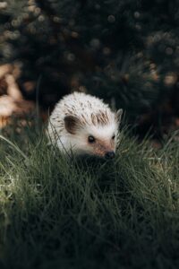
Dermatophytosis (also known as ringworm) is a common fungal infection on the superficial layer of skin caused by one or multiple pathogenic fungi referred to as dermatophytes. This infection is zoonotic and can be transmitted to other animal hosts and humans through contact with an infected organism or contaminated materials (e.g., bedding, fur, and quills).
With exotic pet ownership on the rise, monitoring and attentiveness toward these patients are vital in veterinary care. When caught early, dermatophytosis in hedgehog pets can be addressed prior to the spread to other pets and humans.
We will cover:
Causes of dermatophytosis in hedgehogs
Clinical signs of dermatophytosis in hedgehogs
Diagnosis and treatment dermatophytosis in hedgehogs
Preventative measures for dermatophytosis in hedgehogs
What causes it?
Dermatophytes are a group of diverse fungi that produce enzymes to digest keratin, which is present in skin and hedgehog quills. [2] These fungal pathogens live on the surface of skin, digesting the keratin for nutrients. The high abundance of keratin in hedgehog quills allows for a hospitable environment for dermatophytes to invade and develop.
The most common dermatophyte to cause hedgehog dermatophytosis is Trichophyton erinacei, which has an infection rate between 30-45%. [3] African pygmy hedgehogs (Atelerix albiventris) and Egyptian long-eared hedgehogs (Hemiechinus auratus) are the primary hedgehog species to be infected with T. erinacei; however, T. erinacei can affect all hedgehog species.
T. erinacei is not the only dermatophyte to infect hedgehogs. Other studies have also identified Trichophyton mentagrophytes, Nannizia gypsea [1], and Microsporum spp. [4] as causative agents of dermatophytosis in hedgehogs. With variability in the microorganism causing it, accurate identification is key for a proper treatment plan.
Clinical signs
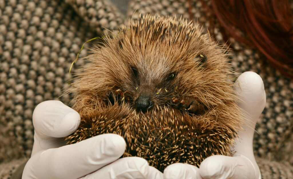
There exists high variability in dermatophytosis symptoms once an exotic pet is infected, ranging from severe alopecia (fur and quill loss) to asymptomatic. [1,4] Crusty, flaky lesions can appear on the skin, which can look scaley or an abnormal texture. Skin lesions can be localized or generalized and are most prominent on the head.
Diagnosis and treatment
Treatment plans for dermatophytosis in hedgehogs have been well documented in recent years. Topical and systemic drug administration have been recorded, both independently and together. However, topical medications may be challenging for the provider and stressful to the animal.
Among the antifungal treatments, itraconazole and terbinafine have been shown to be effective options when given orally with food. Research suggests that terbinafine was more effective than itraconazole, even resulting in a higher elimination rate after 14 days of treatment. [3]
Despite this advancement, the emergence of terbinafine-resistant strains has underscored the need for tailored treatment options. One exotic veterinarian emphasizes the importance of personalized care, stating that it is “essential to choose the most appropriate treatment based on the individual case and the response to therapy.” [3] Close monitoring of the patient’s progress is important to ensure precise targeting of dermatophytes and prevent the development of drug resistance.
 It is vital that pathogenic fungi are targeted effectively, as many species within Trichophyton are morphologically similar and difficult to differentiate. [3] This complexity emphasizes the significance of molecular-based techniques for accurate identification.
The highly conserved nature of the ITS region within fungal genomes allows for unparalleled specificity and reliability. Every fungus has this region in their genome, where it effectively serves as a barcode to identify all fungal species in clinical samples. Molecular techniques such as the MiDOG All-in-One Test allow for all pathogens can be detected and quantified without the long wait time associated with culture media growth.
Because of the specificity of the ITS region, ITS sequencing is regarded as the “gold standard for dermatophyte identification.” [5] It enables a clear distinction between T. erinacei and its close relatives by reading the molecularly unique barcodes. The accurate identification of fungal pathogens using ITS sequencing allows for precision in determining treatment options for each individualized case.
Preventative measures
Diagnosis and treatment of dermatophytosis pose unique challenges to veterinary care providers. Many cases of hedgehog dermatophytosis are asymptomatic or mild, which is difficult to identify when the pet is not observed often.
Early detection is essential for containing the spread of ringworm to other pets and the pet owners. Hedgehog pet owners should visit their veterinarian regularly to ensure that those pets are happy and itch-free.
If you think one of your patients is suffering from dermatophytosis, consider using the MiDOG All-in-One Test today!
References
[1] Le Barzic, C. et.al. (2021). Detection and control of dermatophytosis in wild European hedgehogs (Erinaceus europaeus) admitted to a French wildlife rehabilitation centre. Journal of fungi, 7(2), p. 74. https://doi.org/10.3390/jof7020074.
[2] Khan, A. et.al. (2020). Keratinophilic fungi: Isolation, identification, pathogenicity, characterization, and treatment. New and future developments in microbial biotechnology and bioengineering, p. 29-37. https://doi.org/10.1016/B978-0-12-821006-2.00003-0.
[3] Kottferova, L. et.al. (2023). Hedgehog dermatophytosis: Understanding Trichophyton erinacei infection in pet hedgehogs and its implications for human health. Journal of Fungi (Basel), 9(12), p. 1132. https://doi.org/10.3390/jof9121132.
[4] Ruszkowski, J. et.al. (2021). Hedgehogs as a potential source of zoonotic pathogens—A review and an update of knowledge. Animals (Basel), 11(6), p. 1754. https://doi.org/10.3390/ani11061754.
[5] Kong, F. et.al. (2008). Rapid identification and differentiation of Trichophyton species, based on sequence polymorphisms of the ribosomal internal transcribed spacer regions, by rolling-circle amplification. Journal of Clinical Microbiology, 46(4), p. 1192-1199. https://doi.org/10.1128%2FJCM.02235-07.