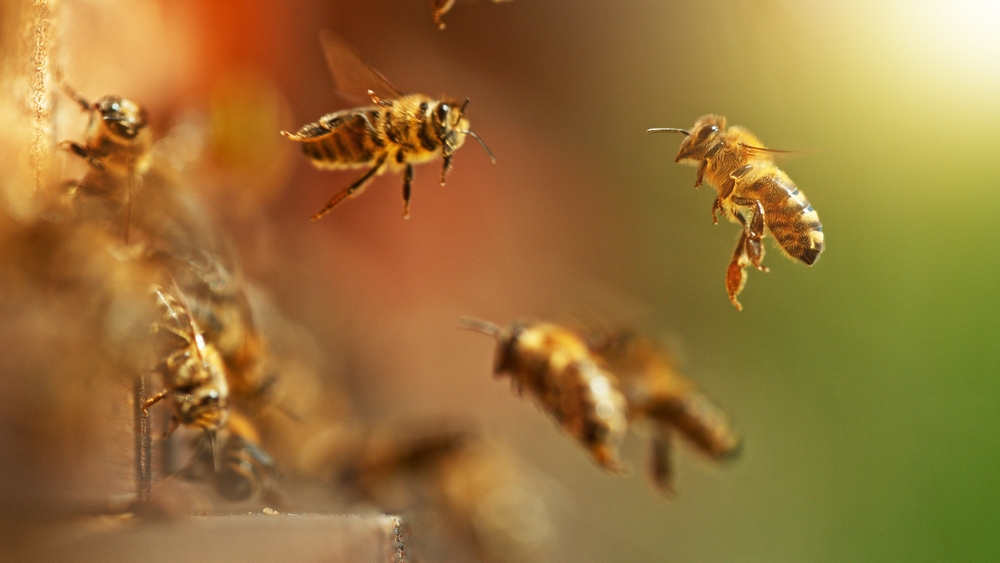
In recent years, the buzz about bees has taken on a more somber tone as the decline in their populations emerges as a global concern. This decline, intricately linked to the delicate balance of ecosystems, highlights the vital role bees play as pollinators. From the hillsides of rural farms to the concrete jungles of urban gardens, bees diligently flit from flower to flower, facilitating the reproduction of plants and the production of fruits and seeds. Yet, these industrious insects face an array of challenges, from habitat loss and pesticide exposure to the insidious threat of pathogens.

Challenges in Bee Health and Ecosystem Impact

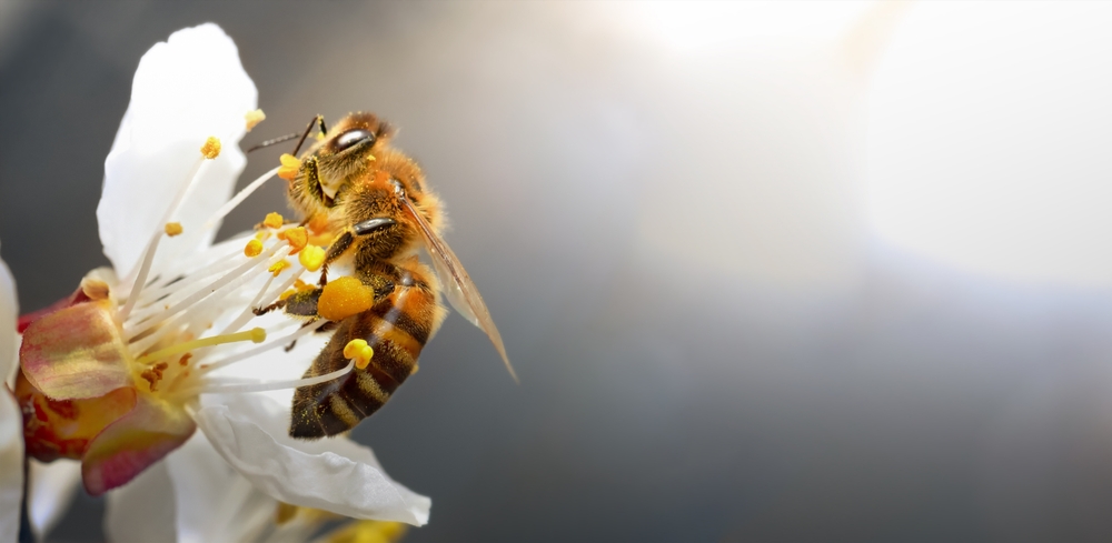
Bees aren’t just important for honey production; they’re essential for maintaining the balance of ecosystems. Their diligent pollination efforts directly impact agricultural productivity and the maintenance of biodiversity. As bee populations dwindle, the ripple effects are felt across the globe, threatening food security and the resilience of ecosystems.

bees and pathogens

Pathogens Threatening Bee Health

Bees, like any other living organisms, are not immune to the threat of disease. They face a barrage of pathogens, including bacteria, fungi, and viruses. Among these, the bacterial culprit Paenibacillus larvae looms large, causing American foulbrood—a devastating disease that leads to larval mortality and hive collapse. Meanwhile, fungal pathogens like Ascosphaera apis trigger chalkbrood disease, stunting larval development. Adding to the woes is the presence of viral infections like Deformed wing virus (DWV), which impairs bee flight and contributes to the mysterious phenomenon of colony collapse disorder.

Emerging Bee Pathogens

Recent research has unveiled new players in the bee health drama, including Lotmaria passim, associated with the Israeli acute paralysis virus (IAPV) complex, and European foulbrood. European foulbrood, caused by the bacterium Melissococcus plutonius, poses a significant threat to bee populations and overall hive health. This bacterial infection primarily affects honeybee larvae, leading to their premature death and weakening of the colony. As infected larvae decompose, they release spores, further contaminating the hive environment and potentially spreading the disease to healthy individuals. The impact of European foulbrood on bee populations can be devastating, as it can result in colony collapse and significant economic losses for beekeepers. According to a study published in the journal “PLoS ONE,” European foulbrood has been identified as a major contributor to honeybee population decline worldwide (Genersch et al., 2006). Efforts to manage and control this disease are crucial to safeguarding bee populations and maintaining the vital role bees play in pollination and ecosystem health. Unfortunately, though, this pathogen is notoriously difficult to diagnose using traditional culture methods, leaving most laboratories unable to diagnose this pathogen. Dr. Joerg Mayer, a full professor in zoological medicine, a board certified veterinarian in exotic animal medicine at the University of Georgia, says “The NGS technique as offered by MiDOG is wonderful because pathogens like Melissococcus plutonius, for example, are difficult to grow and keep alive in the laboratory. A standard laboratory cannot truly examine or even properly diagnose this pathogen in bee hives at this time. But with NGS, we can reliably diagnose it.”

These emerging pathogens pose new challenges to the already delicate balance of bee health management strategies.

Understanding the Bee Microbiome

bee microbiome

Within the buzzing hive lies a hidden world—the bee microbiome—a rich tapestry of microorganisms residing in bee guts and other tissues. These microbiomes include bacteria, fungi, viruses, and other microorganisms, forming complex ecological networks. By studying the composition and function of bee microbiomes, researchers aim to identify beneficial microorganisms that could be harnessed to enhance bee immunity and resilience to pathogens.

This microbial community wields immense influence over bee health and resilience. Deciphering its secrets holds the key to enhancing bee well-being and curbing the impacts of disease.

Among the researchers contributing significantly to the understanding of bee microbiomes is Dr. Joerg Mayer. Dr. Mayer and his team have conducted pioneering studies on the microbial communities associated with bees and their implications for bee health.

Dr. Mayer’s work has shed light on the intricate relationships between bee microbiomes, environmental factors, and disease susceptibility. His research has revealed the diversity of microorganisms inhabiting bees and their dynamic interactions within bee colonies. Through innovative molecular techniques and bioinformatic analyses, Mayer has unraveled the functional roles of specific microbial taxa in bee nutrition, immunity, and pathogen defense.

Key Bacteria Associated with Bee Health

Among the microbial inhabitants of bees, certain bacteria stand out as guardians of health. Lactobacillus and Bifidobacterium, renowned for their probiotic properties, play pivotal roles in bolstering bee immune function and maintaining microbial balance within the hive.

As we delve deeper into the microscopic world of bees, the importance of supporting research efforts becomes ever more apparent. Dr. Mayer and his team at UGA are at the forefront of this critical endeavor, but they can’t do it alone. Your support can make a real difference in safeguarding these essential pollinators and preserving the delicate balance of our ecosystems. Join us in our mission to ensure a thriving future for bees and the planet they call home.

Please consider donating for this great case by clicking below.

Donate!


References:
Engel P, Martinson VG, Moran NA. Functional diversity within the simple gut microbiota of the honey bee. Proc Natl Acad Sci U S A. 2012;109(27):11002-11007.
McFrederick QS, Wcislo WT, Taylor DR, Ishak HD, Dowd SE, Mueller UG. Environment or kin: whence do bees obtain acidophilic bacteria? Mol Ecol. 2012;21(7):1754-1768.
Goulson D, Nicholls E, Botías C, Rotheray EL. Bee declines driven by combined stress from parasites, pesticides, and lack of flowers. Science. 2015;347(6229):1255957.
Kwong WK, Moran NA. Gut microbial communities of social bees. Nat Rev Microbiol. 2016;14(6):374-384.
Raymann K, Moran NA. The role of the gut microbiome in health and disease of adult honey bee workers. Curr Opin Insect Sci. 2018;26:97-104.
Maine Department of Agriculture, Conservation and Forestry. “Diseases of Honey Bees.” https://www.maine.gov/dacf/php/apiary/documents/factsheets/diseases.pdf
Nazzi, Francesco, and Jay D. Evans. “Ecology and Pathophysiology of the Honey Bee Gut Pathogen Nosema ceranae.” Journal of Invertebrate Pathology, vol. 103, no. Supplement 1, 2010, pp. S10–S19. https://www.ncbi.nlm.nih.gov/pmc/articles/PMC7783639/#:~:text=Moreover%2C%20most%20common%20viruses%20that,bee%20paralysis%20virus%20(SBPV)%2C.
Genersch, E., Forsgren, E., Pentikäinen, J., Ashiralieva, A., Rauch, S., Kilwinski, J., & Fries, I. (2006). Reclassification of Paenibacillus larvae subsp. pulvifaciens and Paenibacillus larvae subsp. larvae as Paenibacillus larvae without subspecies differentiation. PLoS ONE, 1(1), e45. https://doi.org/10.1371/journal.pone.0000045


Categories: Gut Microbiome, Insects

animal diagnostics

Validated by Veterinarians

“Clients expect their veterinarians to stay up to date on all matters that affect the health and well being of their non-human family members. The current technique that we have used to determine the presence and antibiotic sensitivity of organisms causing disease in our pets is over a century old.

With the emergence of dangerous antibacterial resistance, it is critical that veterinarians are able to offer laser focused diagnostics and treatment. MiDog enables us to offer care that exceeds the typical standard of care.”

Bernadine Cruz, DVM, Laguna Hills Animal Hospital Laguna Woods, CA

“I love the absolute abundance and comparing the fungal with bacterial infection. I do not worry as much about getting a false negative urinary infection reading as I do with traditional urine cultures. Several times the same urine would culture negative but MiDOG would detect pathogens.”

Michael Morgan, DVMQuail Animal Hospital, Tustin, CA

“The MiDOG All-in-One Test is amazing, I would use it instead of culture and sensitivity.  Such rapid and detailed results, I will reach for MiDOG before culture next time!

Thank you very much MiDOG, for sharing the opportunity to try your technology.”

Martha Smith-Blackmore, DVM, President of Forensic Veterinary Investigations, LLC – Boston, MA

The MiDOG All-in-One Microbial Test is our new gold standard of pathogen identification. The results are so accurate and valuable – especially with assessing both bacterial and fungal infections with the same sample.

Thank you MiDOG!”

Kathy Wentworth, DVM, Diplomate ABVP Canine and Feline Practice – PetPoint Medical Center, Irvine, CA

“The MiDOG staff was extremely helpful and supportive.”

Cathy Curtis, DVM – London, UK

“I have had great results using the MiDOG® Test. Compared to traditional culture tests, I am better able to target the treatment for dogs because the MiDOG® Test is so sensitive that it identifies all pathogens including bacteria and fungi, as well as antibiotic sensitivity.

The cost and turnaround time are about the same as a culture test, but I get much more data. The test has great performance and I believe the NGS technology will be a game changer for veterinarians treating dogs with lesions or other infections.”

Michael Kavanagh, DVM, Practice owner – Saddleback Animal Hospital, Tustin, CA

“It’s helpful to have an NGS spectrum because it gives you a broader insight of what’s happening and what might be going on.”

Richard Harvey BVSc DVD DipECVD PhD FRSB FRCVS – European Specialist in Veterinary Dermatology – Head of Dermatology, Willows Veterinary Centre & Referral Service – Solihull, England UK

“I have been using MiDog for over 4 years now and exclusively as my test of choice for all cultures for 3 years.  It is so great to submit a culture and feel confident there will be a result when it comes back, especially for urine cultures.  The reports were intimidating at first because they contain so much information.  After the first few, I am now quickly able to glance over it and pick out the highlights.  I can then come back later and pour over all the details.  I have been extremely pleased with my patients’ results using the test as well.  I don’t envision ever going back to traditional culture and susceptibilities again.”

Brian M. Urmson, DVM, Columbiana Veterinary Associates

“As an exotic veterinarian, there are numerous tests we have to consider to check specific bacterial and fungal organisms based on the species. MiDOG eliminates the need for many of these separate samples and provides definitive results quickly to help us treat our patients more efficiently and effectively. The lab is wonderful to work with and has never rejected our samples- they even processed a lizard toe we amputated and determined the cause of skin infection.”

Dr. Melissa Giese, Chicago Exotics Animal Hospital

“MiDOG’s diagnostic approach offers the unique ability to identify pathogens that evade traditional culture and sensitivity testing. I have found that adding a molecular based testing approach in the form of Next Generation Sequencing (NGS) from MiDOG to my routine diagnostic cultures can be extremely helpful in the identification and diagnosis of uncommon pathogens in veterinary medicine.”

Dr. Wayne Rosenkrantz, Animal Dermatology Clinic – Tustin

“She [Dr. Krumbeck] really did a great job of making complicated concepts accessible and demonstrating the value of your services. I’m really looking forward to working with MiDOG on my research project!”

Dr. Yaicha Peters, Animal Dermatology Clinic – San Diego

“As a proud collaborator with MiDOG, I deeply appreciate their dedication to fostering partnerships between industry and veterinary experts. Their commitment to enhancing diagnostic quality for veterinarians is commendable. In my experience, their support has been invaluable, earning them a ‘Double A+, Triple Star’ rating. Their assistance has been faultless, contributing significantly to the success of my projects and studies. I eagerly anticipate our continued collaboration.”

Dr. Richard Harvey, BVSc DVD DipECVD PhD FRSB FRCVS; European Specialist in Veterinary Dermatology

“We’re seeing that, if we prescribe too many antibiotics or they’re taken too frequently, animals are developing inappropriate or pathogenic strains of bacteria. We’re also seeing that our antibiotics are just not working against them anymore… It’s a good example of why we need better diagnostic testing, like MiDOG, so that we’re selecting the correct antibiotic every time our patients have an infection.”

Dr. Alissa Rexo, DVM, CVA, DACVD, Mid-Atlantic Veterinary Dermatology

“The NGS technique as offered by MiDOG is wonderful because pathogens like Melissococcus plutonius, for example, are difficult to grow and keep alive in the laboratory. A standard laboratory cannot truly examine or even properly diagnose this pathogen in bee hives at this time. But with NGS, we can reliably diagnose it.”

Dr. Joerg Mayer, Entomologist and Microbiologist at the University of Georgia

“For me, as a clinician and as a researcher, I see the immense value in the product [the All-in-One Test]. I have had great success using MiDOG clinically, including identifying Mycoplasma in a 24 year old pigeon, a Nannizziopsis spp in a ball python from a large pet distributor, Mycoplasma and Fusobacterium necrophorum co-infection in a peacock, and to identify an abnormal gut GI in a technician’s dog that tested negative for everything else (but we were able to establish what was abnormal, and work toward fixing it).”

Dr. Jeremy Rayl – Veterinarian, Block House Creek Animal Hospital, Cedar Park, Texas

“With MiDog results I not only receive a list of organisms detected in the sample, but also cell counts which allow me to target the most abundant pathogens or the most pathogenic. The identification of antimicrobial resistance genes is also helpful in selecting an appropriate antibiotic. Another benefit is that sample handling is less prone to risk of overgrowth and is stable at room temperature. Easy and informative. A great tool in our diagnostic toolbox!!”

Lauren Palmer DVM, MPH, Marine mammal Care Center Los Angeles